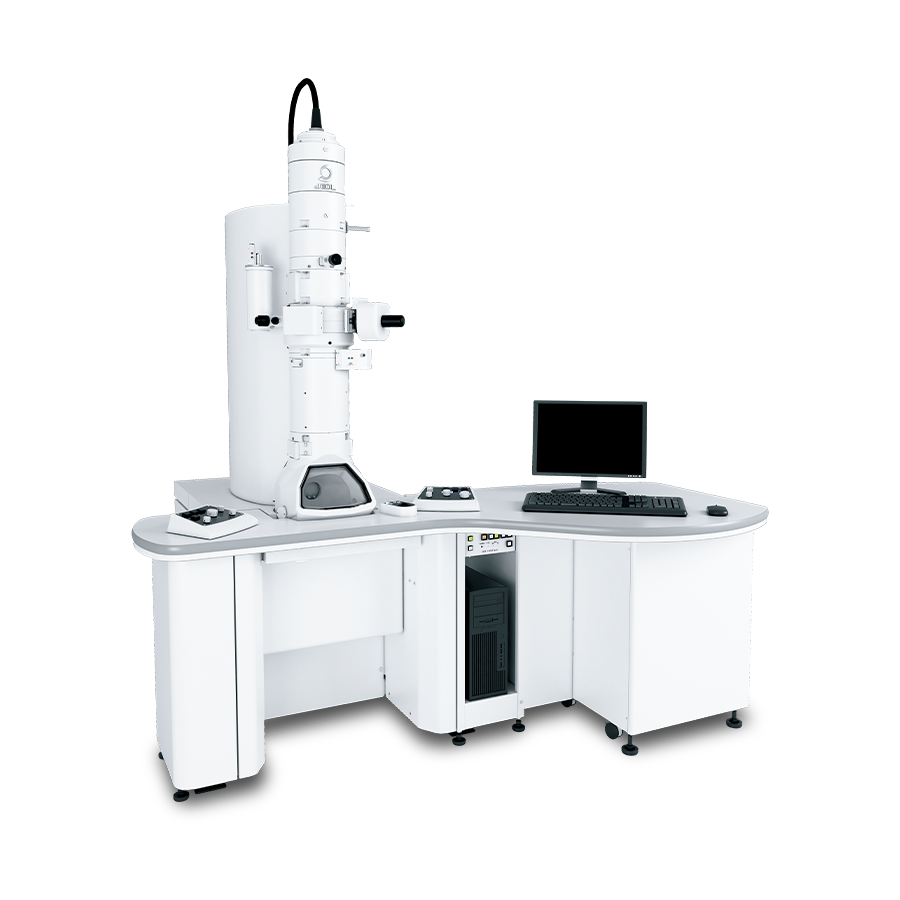
【DISCONTINUED】JEM-1400Flash
Electron Microscope
DISCONTINUED
This product is no longer available.
If you would like to know the latest information about your preferred product or to find out more about alternatives, please click on the link below. We hope you will continue to use our products.
The JEM-1400Flash is used in a wide range of fields, such as biology, nanotechnology, polymer, and advanced materials. In the observation of biological specimens including macro-molecular materials, medicines, pathological sections and viruses, usually the entire view of tissues, structures, target locations and observation area are first confirmed at low magnification, and then fine structures of interest are carefully studied at high magnification. To smoothly proceed to this series of observation, recent demands for easier observation steps to acquire higher-throughput image data are increasing. To meet those needs, a new 120 kV electron microscope “JEM-1400Flash” is equipped with a high-sensitivity sCMOS camera, an ultra-wide area montage system, and an OM (optical microscope) image linkage function.
Features
High-sensitivity sCMOS camera, "Matataki Flash"
"Matataki Flash", JEOL’s innovative high-sensitivity sCMOS camera, dramatically reduces the readout noise while possessing high frame rate. This powerful feature enables high-throughput acquisition of sharp TEM images with extremely low-noise.
New function: Ultra-wide area montage system, Limitless Panorama (LLP)
In addition to the conventional electromagnetic image shift, the JEM-1400Flash comes with a montage system capable of utilizing stage drive for the field shift. This new system allows for simple capture of a montage panorama image over a limitless wide area. Thus, an ultra-wide area, high pixel-resolution image is obtainable, which is comparable to the image taken by conventional photo film.
The combined use with "Matataki Flash" sCMOS camera enables automatic acquisition of "Limitless Panorama" pictures with no limitation of the number of pixels.
New function: OM image linkage function, Picture Overlay
A digital image acquired with an OM can be overlaid on a TEM image. Since an observation area can be searched on the overlaid image, high resolution observation of a fluorescence site is easily made on the TEM image.
New exterior design: "Pure white" and new user interface
JEOL products’ brand color of "pure white" is newly adopted, leading to a sophisticated exterior design of the JEM-1400Flash. New designs are also employed for (1) the control panel, with an LED lamp for easy-to-view operation, (2) a small work table enabling compact layout of the user controls, and (3) a new smart black Graphical User Interface.
Specifications
| Resolution | 0.2 nm (HC) 0.14 nm(HR) |
|---|---|
| Accelerating voltage | 10 to 120 kV |
| Magnification | ×10 to ×1,200,000 (HC), ×10 to ×1,500,000 (HR) |
| Max. tilt angle | ±70° *With the optional high tilt specimen holder |
| Number of specimens to load | Up to 4 *With the optional specimen quartet holder |
| Vacuum system | TMP |
Catalogue Download
JEM-1400Flash Electron Microscope
Application
Application JEM-1400Flash
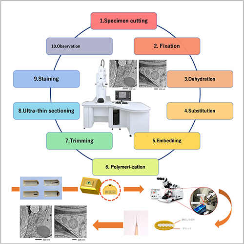
Observation of biological specimen by JEM-1400Flash ー Flow from sample preparation to observation ー
High resolution TEM image by JEM-1400Flash
Ultrawide transmission electron microscopy image of a mouse kidney
Wide Area Observation by Using SiN Window Chip
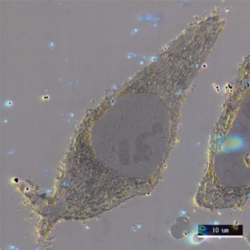
Observation of TOM20 localization in HeLa cell
Gold particles eaten by paramecium
Identification of fine particles emitting fluorescence in toner
Specimen Preparation Method
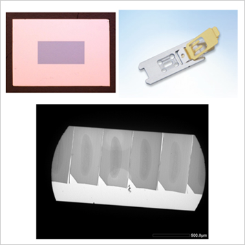
SiN Window Chip and its applications
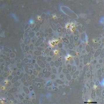
Wide Area Observation by Using SiN Window Chip
An ultra wide highly precise image is obtained by using SiN Window Chip and the automatic montage system "Limitless Panorama (LLP)" function.
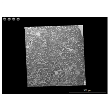
Ultrawide transmission electron microscopy image of a mouse kidney
Ultrathin sections of a mouse kidney mounted on a SiN Window Chip were imaged using the Limitless Panorama (LLP) function, an automated montage system.
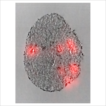
Identification of fine particles emitting fluorescence in toner
Using the "on chip CLEM" which enables overlaying images with high precision, we are able to easily identify fine particles that emitted fluorescence and perform detailed TEM observations.
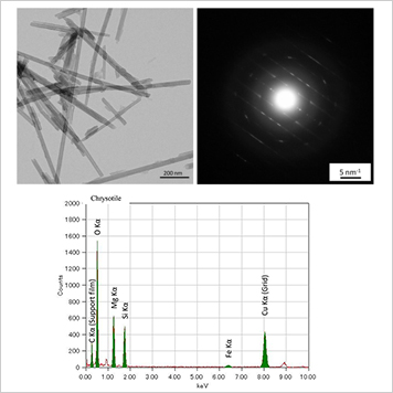
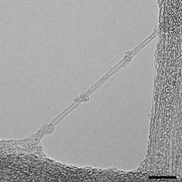
Asbestos / High Resolution TEM Image
Related Products
Related Products
SightSKY Camera (EM-04500SKY) High sensitivity, Low noise fiber coupling CMOS camera
・High-sensitivity, low-noise 19 M pixel CMOS sensor enables clearer imaging with fine specimen details observable even at low electron doses.
・Its global shutter and high frame rate (58 fps/full pixel mode) enable image series acquisitions with less artifacts during dynamic observation.
・"SightX" camera system control software provides user-friendly operations.
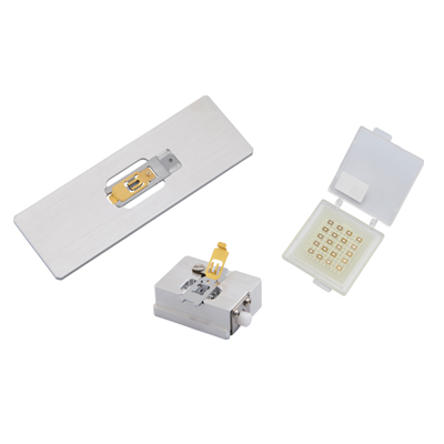
SiN Window Chip
The high-strength SiN film enables us to observe a serially large area of a millimeter in size. It is also ideal for observation of serially sliced sections because there is no invisible area that is caused by conventional TEM grids. The dedicated retainer makes it easy to perform Correlative Light and Electron Microscopy (CLEM).
More Info
Are you a medical professional or personnel engaged in medical care?
No
Please be reminded that these pages are not intended to provide the general public with information about the products.