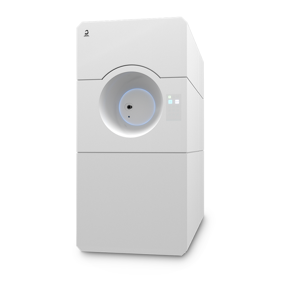
SightSKY™ Series
High-Sensitivity, Low-Noise
Fiber-Coupling CMOS Camera
・High-sensitivity, low-noise 19 M pixel CMOS sensor enables imaging of fine specimen detail and obtains high signal to noise images even at low electron doses.
・A Global shutter and high frame rate (58 fps/full pixel mode) enable image series acquisitions with less artifacts during in-situ dynamic observation studies.
・The SightSKY™ camera system offers integration with JEOL’s FEMTUS™ Integrated Analysis Platform, providing user-friendly operation and data acquisition.
Features
High-resolution TEM image of silicon nitride (Si3N4)
Digital zoom ensures clear, detailed images even when viewing at atomic resolution.
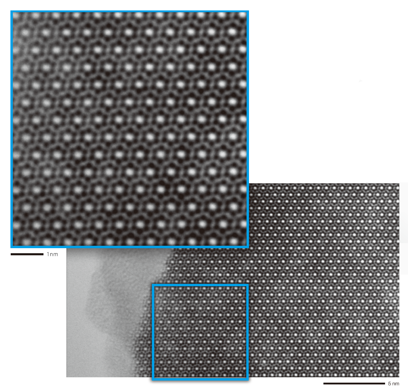
Instrument: JEM-2100Plus
Accelerating voltage: 200 kV
High-resolution TEM image and electron diffraction pattern of Al72Fe24Ni4 decagonal quasi-crystal
Digital Zoom enables observation of atomic structure showing quasi-periodicity, such as shown when imaging quasi-crystals.
Moreover, the high dynamic range offers excellent electron diffraction pattern contrast, from the high-intensity direct spot 0 order spot to weaker-intensity reflections.
Specimen courtesy of:
Dr. Kenji Hiraga, Professor Emeritus,
Institute for Materials Research, Tohoku University
Dr. Kunio Yubuta, Specially Appointed Professor,
Institute for Aqua Regeneration, Shinshu University
Upper right triangle shows Log display.
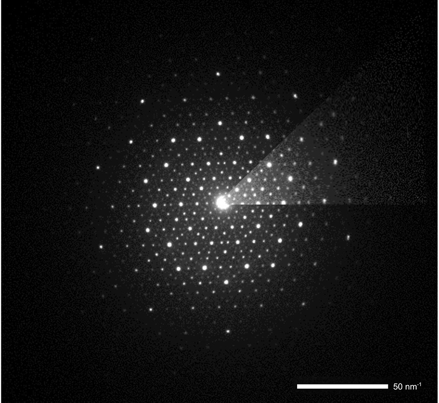
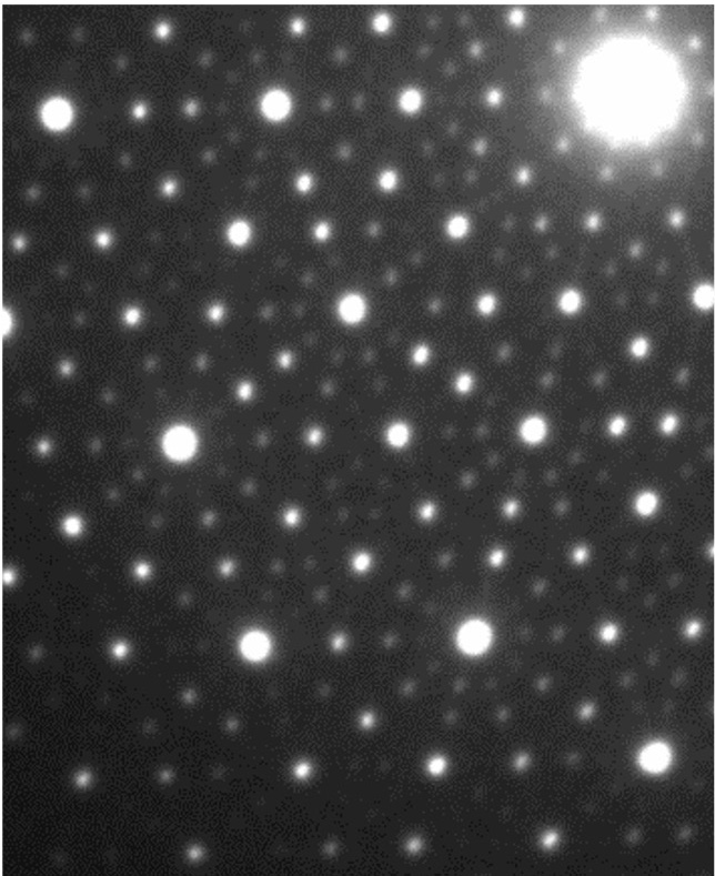
Instrument: JEM-2100Plus,
Accelerating voltage: 200 kV
TEM images of polycrystalline silicon
Fine structures, such as twin textures of polycrystalline silicon, can be observed with high contrast.
BF-TEM
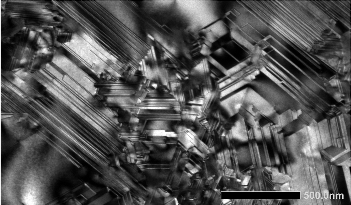
DF-TEM
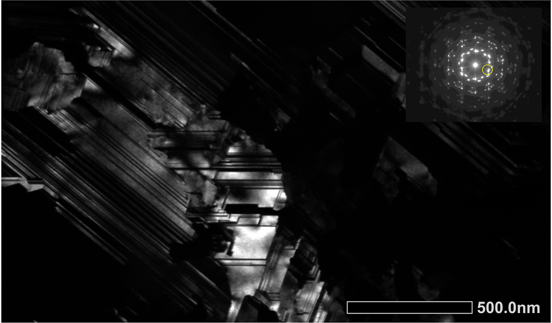
A dark-field image is formed with the indicated diffraction spot.
Instrument: JEM-2100Plus
Accelerating voltage: 200 kV
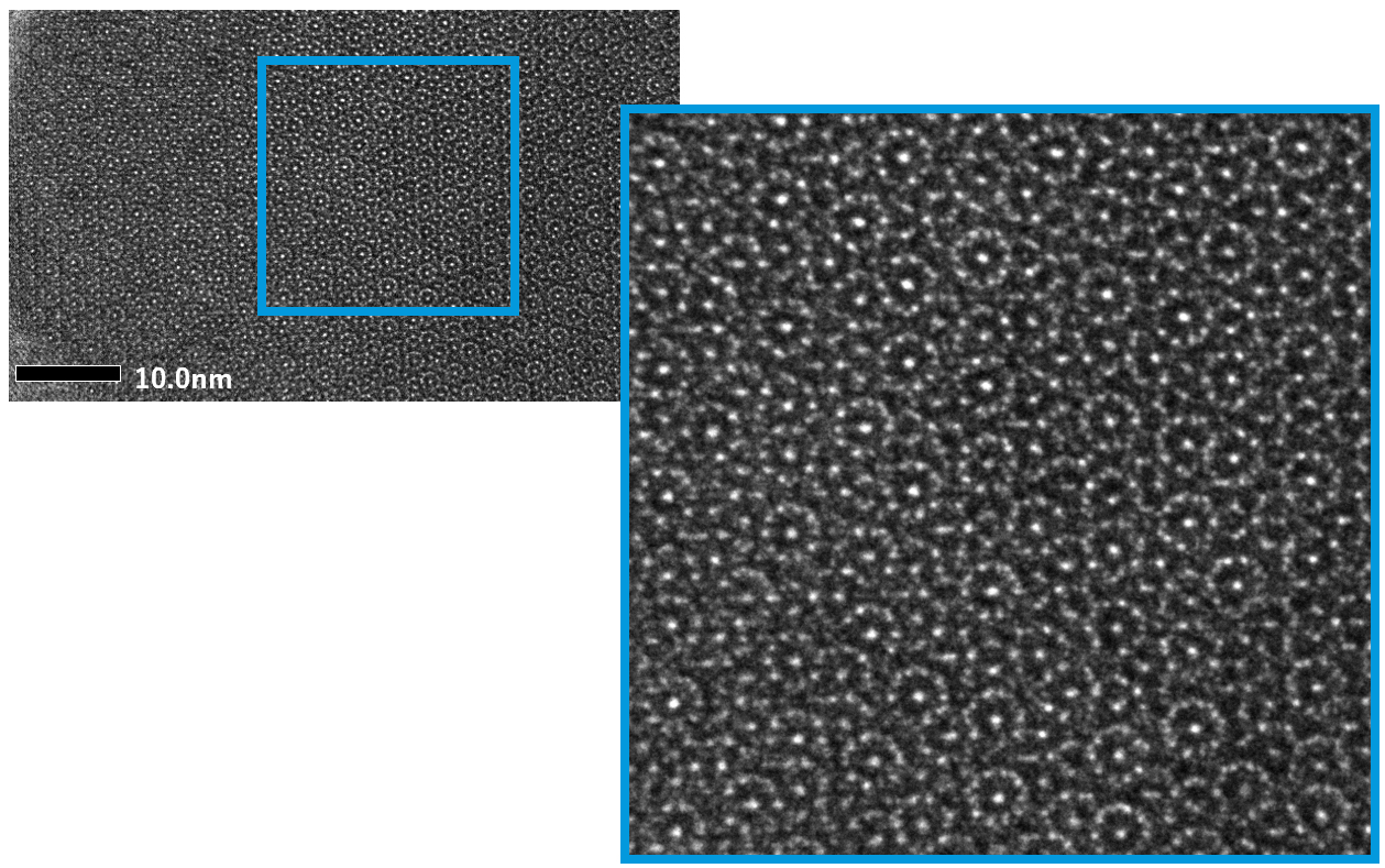
TEM images of MoSi2-Mo5Si3 eutectic alloy
The MoSi2-Mo5Si3 eutectic structure and MoSi2 and Mo5Si3 lattice images can be clearly observed.
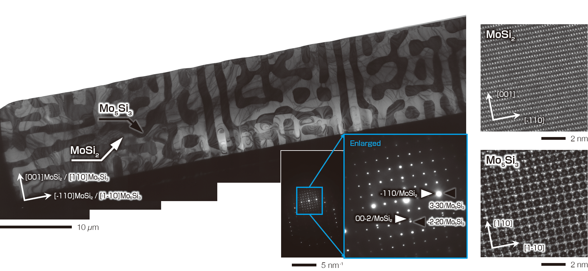
Instrument: JEM-F200,
Accelerating voltage: 200 kV,
Specimen preparation: JIB-PS500i
Specimen courtesy of:
Dr. Haruyuki Inui and Dr. Kyosuke Kishida, Professors,
Graduate School of Engineering, Kyoto University
TEM image of polyethylene/polypropylene resin
Digital Zoom enables clear observation of lamella structures
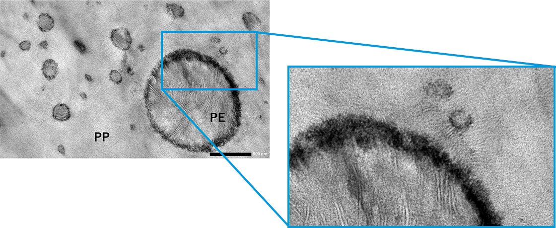
Specimen preparation: Microtome,
Instrument: JEM-2100Plus,
Accelerating voltage: 80 kV
TEM image of Liposome lipid bilayer membrane
The Liposome lipid bilayer membrane can be clearly distinguished.
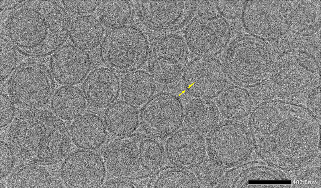
Specimen preparation: ice-embedding,
Instrument: JEM-F200 (CR),
Accelerating voltage: 200 kV
TEM image of mouse kidney
The renal glomerulus fine structure can be imaged with high contrast allowing for accurate clinical diagnosis.
In particular, important structures in renal biopsy observations, such as the basement membrane and foot processes (arrow), can be clearly imaged at low and high magnification.
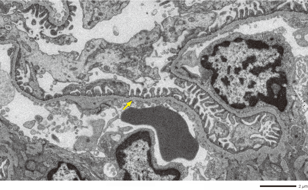
Specimen preparation: Microtome,
Instrument: JEM-120i,
Accelerating voltage: 120 kV
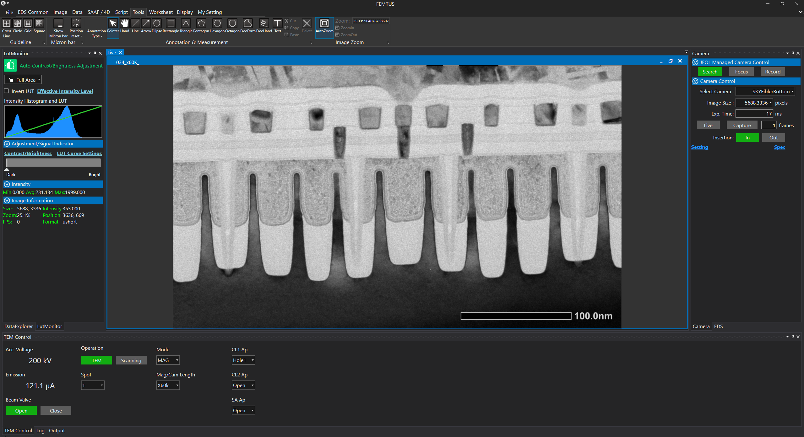
Smart Control with JEOL’s FEMTUS™ Integrated Analysis Platform
The camera system is integrated into JEOL’s FEMTUS™ Integrated Analysis Platform and providing user-friendly operation.
Specifications
| SightSKY™ (EM-04500SKY) |
SightSKY™-R (EM-Z24114TSKYR) |
|
| Retraction mechanism |
Not installed | Installed |
| Applicable accelerating voltage | 200 kV or lower | 300 kV or lower |
| Effective pixels | 19 M pixels (5,688 x 3,336 pixels) | |
| Pixel size | 6.4 μm × 6.4 μm | |
| Frame rate | 58 fps / full pixel mode | |
| Shutter type | Global shutter | |
| Mounting position | Bottom mount (below viewing chamber) |
Pre-filter camera housing (EM-Z211231PCHSG) |
| Binning | x1, x2, x4 | |
| File format | TIFF, Bitmap, JPEG | |
| Recording mode | Image, stack frame (up to 256 frames) | |
| Dynamic range | 16 bit | |
| SightSKY™ (EM-04500SKY) |
SightSKY™-R (EM-Z24114TSKYR) |
Control Software | |||
| FEMTUS™ | SightX | TEM Center | |||
| JEM-120i | ✔︎ |
✔︎ |
|||
| JEM-1400 | ✔︎ |
✔︎ |
|||
| JEM-2100Plus | ✔︎ |
✔︎ |
|||
| JEM-2200FS | ✔︎ |
✔︎ |
|||
| JEM-F200 | ✔︎ |
✔︎ |
✔︎ |
✔︎ |
|
| JEM-ARM200F | ✔︎ |
✔︎ |
✔︎ |
||
| JEM-ACE200F | ✔︎ |
✔︎ |
✔︎ |
||
| JEM-ARM300F | ✔︎ |
✔︎ |
✔︎ |
||
| JEM-3300 | ✔︎ |
✔︎ |
|||
Catalogue Download
SightSKY (EM-04500SKY) High sensitivity, Low noise fiber coupling CMOS camera
Related Products
Related Products
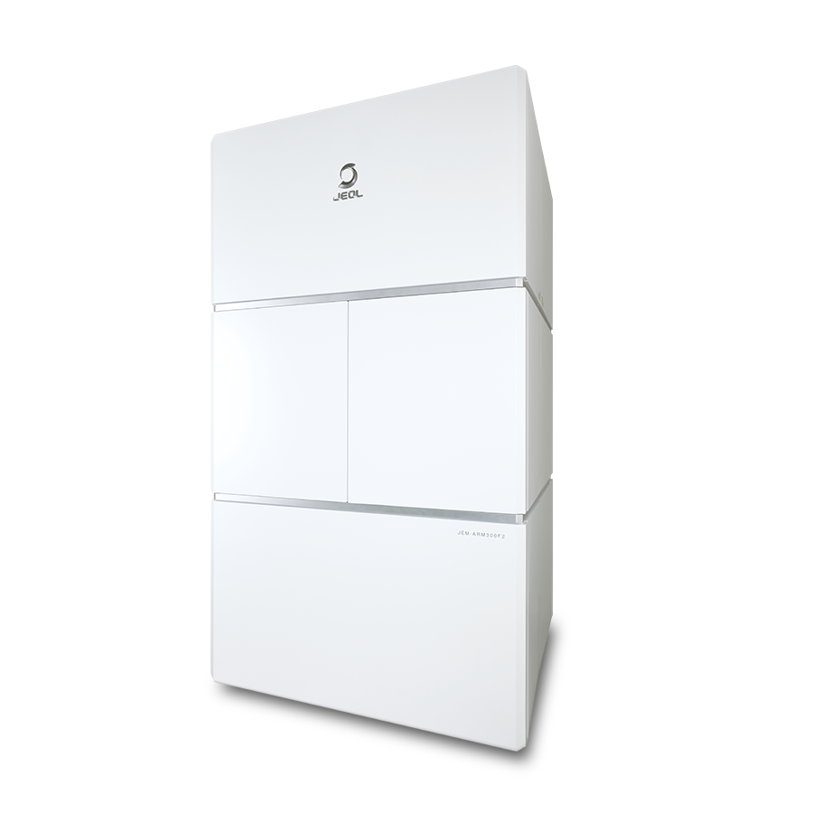
JEM-ARM300F2 GRAND ARM™2 Atomic Resolution Analytical Microscope
The "GRAND ARM™2" has been upgraded.
This "GRAND ARM™2" enables observation at ultrahigh spatial resolution with highly sensitive analysis over a wide range of accelerating voltages.
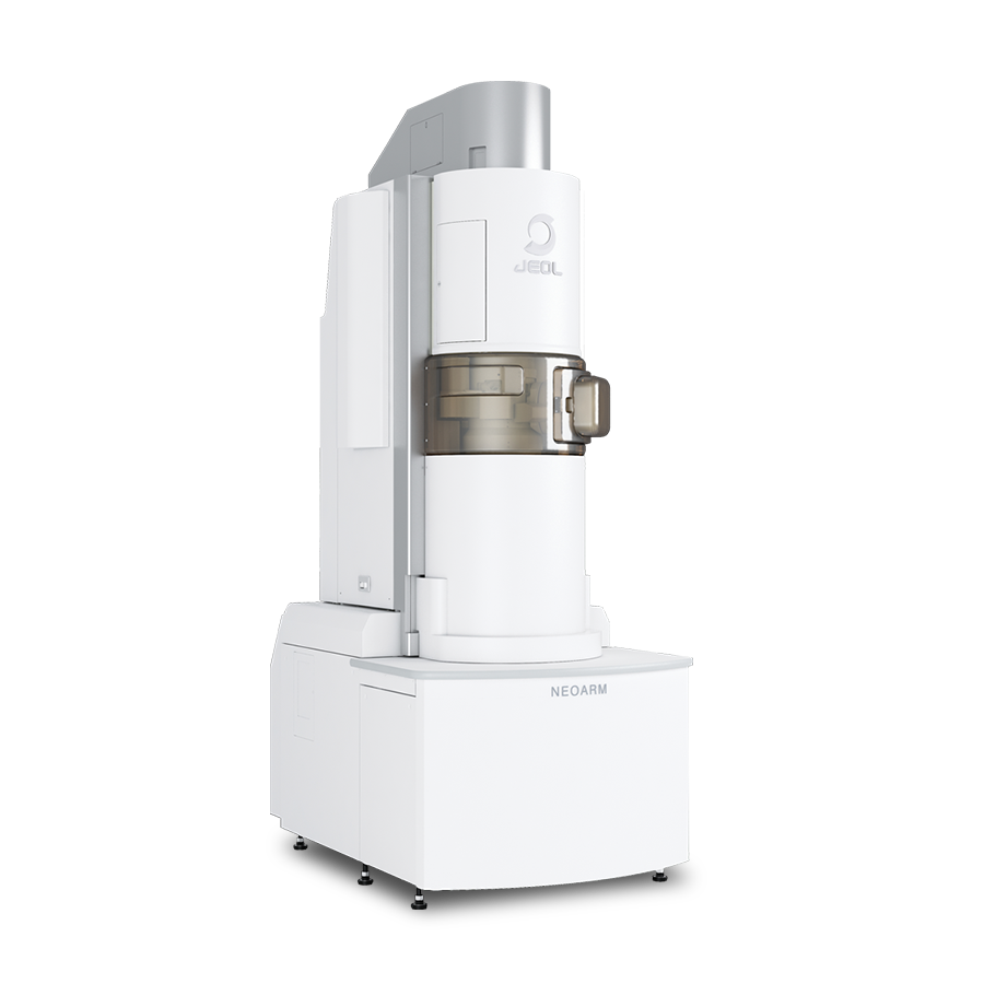
JEM-ARM200F NEOARM Atomic Resolution Analytical Electron Microscope
"NEOARM" / JEM-ARM200F comes with JEOL’s unique cold field emission gun (Cold FEG) and a new Cs corrector (ASCOR) that compensates for higher order aberrations. The combination of a Cold FEG and ASCOR enables atomic-resolution imaging at not only 200 kV accelerating voltage, but also a low voltage of 30 kV."NEOARM" is also equipped with an automated aberration correction system that incorporates JEOL’s new aberration correction algorithm for automatic fast and precise aberration correction. This system enables higher-throughput atomic-resolution imaging.
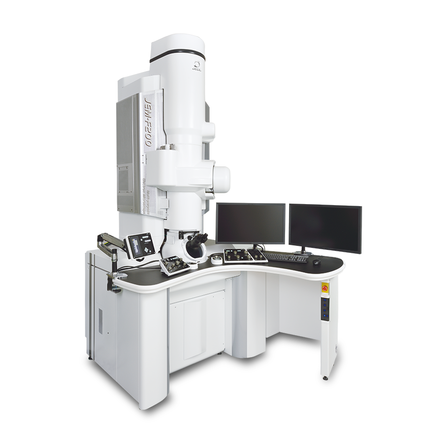
JEM-F200 Multi-purpose Electron Microscope
The JEM-F200 is a field emission transmission electron microscope, which features higher spatial resolution and analytical performance coupled with intuitive user interface for multi-purpose operation.
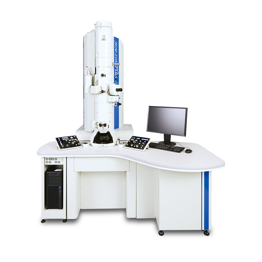
JEM-2100Plus Electron Microscope
The JEM-2100Plus is a multi purpose transmission electron microscope, which combines the proven JEM-2100 optic system with an advanced control system for enhanced ease of operation. Achieving superior performance through intuitive operation, the JEM-2100Plus provides solutions to a wide range of applications from materials science to medical/biological studies.
JEM-120i Electron Microscope
Transmission Electron Microscopes(TEM) with 120kV accelerating voltage are widely used in soft material fields such as biology and polymer. We newly developed JEM-120i with the concept of "Compact", "Easy To Use", and "Expandable". With the new external appearance, this instrument has evolved into a useful tool that anyone can use easily, from operation to maintenance.
More Info
Are you a medical professional or personnel engaged in medical care?
No
Please be reminded that these pages are not intended to provide the general public with information about the products.