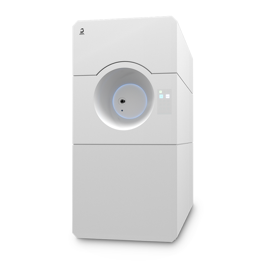
JEM-120i
Electron Microscope
It is a Useful Tool for Every User!
Transmission Electron Microscopes(TEM) with 120kV accelerating voltage are widely used in soft material fields such as biology and polymer. We newly developed JEM-120i with the concept of "Compact", "Easy To Use", and "Expandable". With the new external appearance, this instrument has evolved into a useful tool that anyone can use easily, from operation to maintenance.

Features
Compact
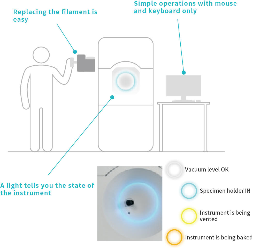
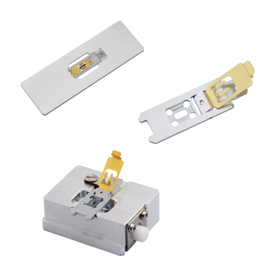
With the drastic reduction in size, the JEM-120i has a filament replacing position and specimen holder insertion position lower than existing instruments. A newly developed cartridge type filament unit helps make filament replacement easy and safe for anyone.
The instrument has an LED light on its front and its color changes depending on the state of the TEM. You can determine its operating state at a distance.
Simple Operation
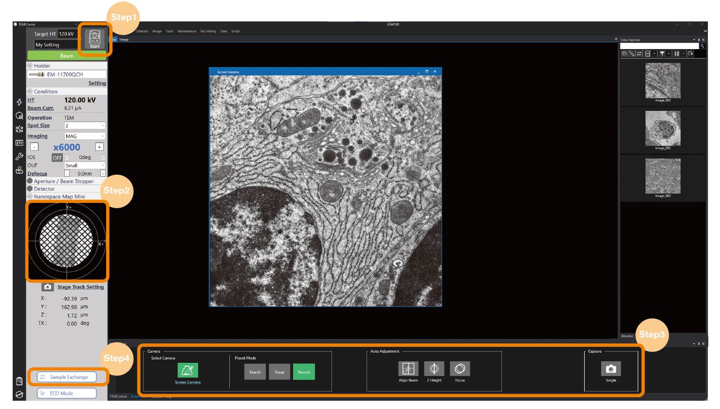
Specimen observation with simple operation
It takes only 4 steps from loading a specimen to completing observation. After inserting the specimen holder, clicking the Start Button automatically performs observation preparation operations such as voltage increase and emission start. A wide area image is captured at the same time. So, clicking the target field will move the stage to the clicked position.
Since standard "Butler mode" assists data acquisition, even a beginner can capture data easily. When observation completes, you can exchange the sample holder while keeping the emission on.
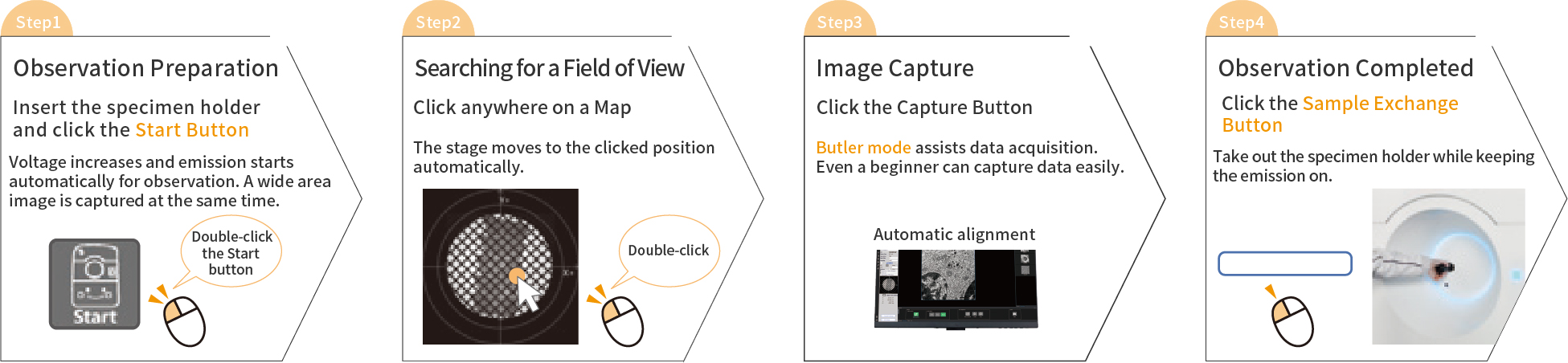
Specimen observation in 4 steps
Seamless observation without switching magnification mode
The JEM-120i is equipped with an enhanced TEM control system and fully automated apertures, eliminating the need for switching magnification modes and selecting an aperture. Observation operations can be performed more smoothly than with previous models.
Expandability
The JEM -120i supports a variety of retrofits. It also meets the need for additional functions after purchase.
Video
Related Link
-A Useful Tool for Every User! - New Electron Microscope JEM-120i Released
The reason the 75th year TEM looks like a smart home appliance
Specifications
| Resolution | 0.2 nm (HC)※1, 0.14 nm (HR)※2 |
|---|---|
| Accelerating voltage | 20-120 kV |
| Magnification | 50-1,200,000 (HC)※1, 50-1,500,000 (HR)※2 |
| Standard camera | NEOView Camera 4 M pixel, 30 fps |
| Optional camera | SightSKY 19 M pixel, 58 fps Cameras by other company can be mounted. |
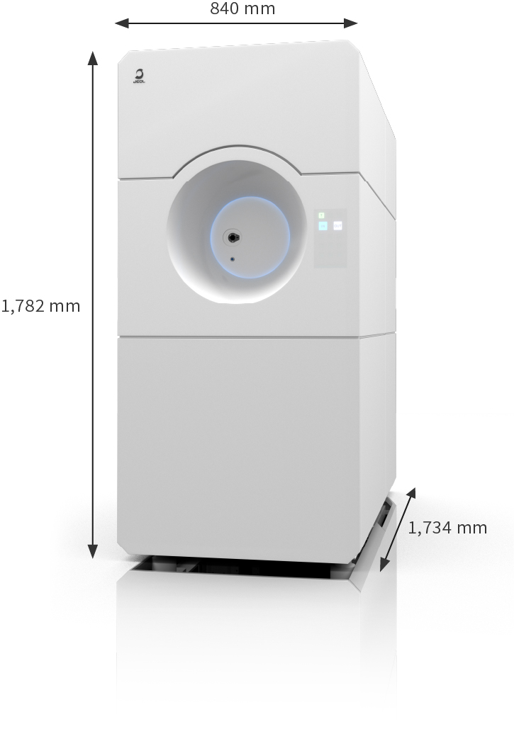
| Main unit dimensions | W 840 mm/ D 1,734 mm/ H 1,782 mm |
1 (HC) : High contrast configuration
2 (HR) : High resolution configuration
Catalogue Download
Application
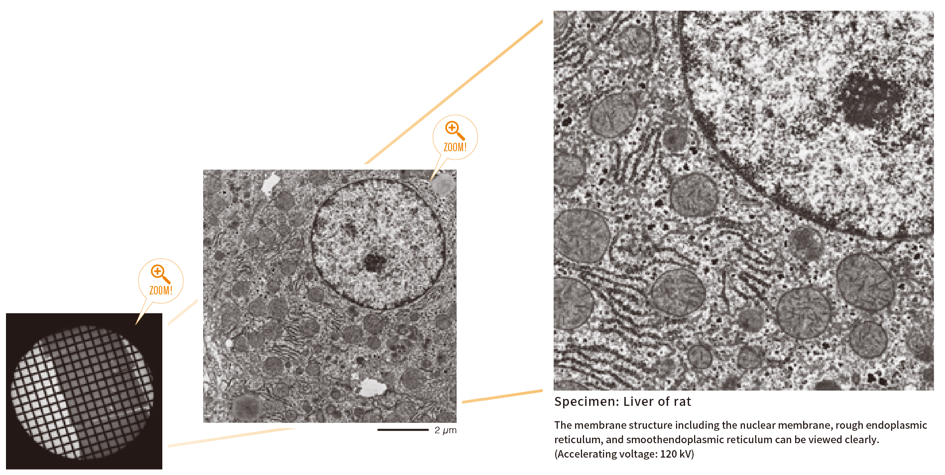
Observation of biological specimen by JEM-1400Flash ー Flow from sample preparation to observation ー
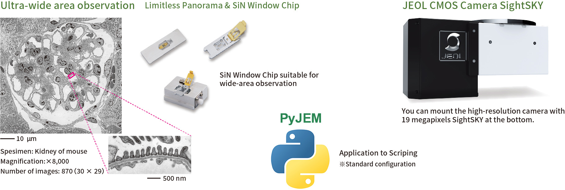
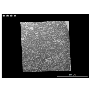
Ultrawide transmission electron microscopy image of a mouse kidney
Wide Area Observation by Using SiN Window Chip
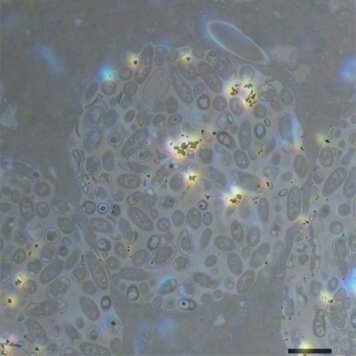
Gold particles eaten by paramecium
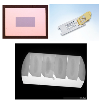
SiN Window Chip and its applications
Wide Area Observation by Using SiN Window Chip
An ultra wide highly precise image is obtained by using SiN Window Chip and the automatic montage system "Limitless Panorama (LLP)" function.
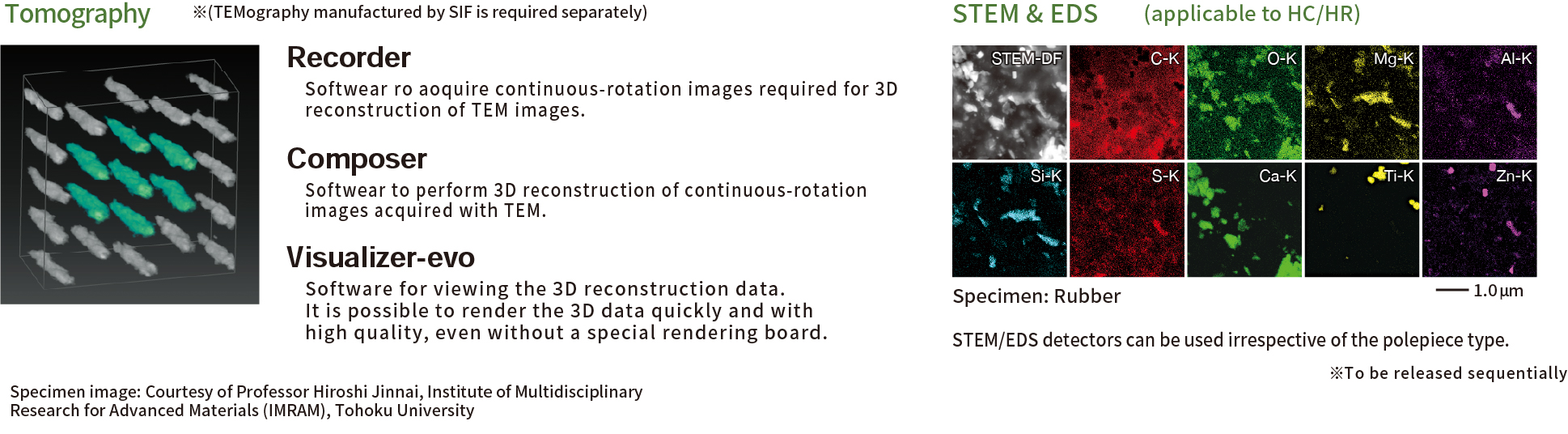
Related Products
SightSKY (EM-04500SKY) High sensitivity, Low noise fiber coupling CMOS camera
High-sensitivity, low-noise 19 M pixel CMOS sensor enables clearer imaging with fine specimen details observable even at low electron doses.
Its global shutter and high frame rate (58 fps/full pixel mode) enable image series acquisitions with less artifacts during dynamic observation.
"SightX" camera system control software provides user-friendly operations.
SiN Window Chip
The high-strength SiN film enables us to observe a serially large area of a millimeter in size. It is also ideal for observation of serially sliced sections because there is no invisible area that is caused by conventional TEM grids. The dedicated retainer makes it easy to perform Correlative Light and Electron Microscopy (CLEM).
More Info
Are you a medical professional or personnel engaged in medical care?
No
Please be reminded that these pages are not intended to provide the general public with information about the products.