Ultrawide transmission electron microscopy image of a mouse kidney
EM2020-10E
Ultrathin sections of a mouse kidney mounted on a SiN Window Chip were imaged using the Limitless Panorama (LLP) function, an automated montage system. Sections mounted on a flat, grid-bar free SiN Window Chip allow us to view the entire area without any wrinkles. To record this wide field of view in high resolution, automatic montage imaging (96 × 90 images) was carried out using LLP at a pixel size of approximately 5.6 nm/pixel. The images thus obtained have approximately 20 billion pixels, allowing us to observe the entire kidney cortex region (distribution of glomeruli and the network of tubules) while maintaining a resolution that allows us to observe the basement membrane structure of the glomerulus.
The following image have been processed from wide-area images taken by LLP and can be viewed with a Web browser. Clicking on the image will open a new tab for viewing the wide area image.
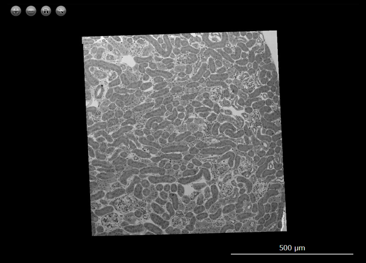
Sample : Mouse kidney
Imaging Device : JEM-1400 / Matataki Flash Camera (2,048 x 2,048 pixels)
Image acquisition area : H 800 μm x W 760 μm with 8,640 (H 96 x W 90) images
Pixel size of the image : 5.6 nm/pixel
Total number of the pixels : 20 Gigapixel
Acceleration Voltage : 80 kV
Data provided by
RIKEN Center for Integrative Medical Sciences
Department of Nephrology, Graduate School of Medicine, Kyoto University
Dr. Oguchi
- Please see the PDF file for the additional information.
Another window opens when you click. 
PDF 718KB
Are you a medical professional or personnel engaged in medical care?
No
Please be reminded that these pages are not intended to provide the general public with information about the products.