

Clinical / Pathological Tests
Both clinical tests and pathological tests play an important role in medicine.
A clinical test is used to understand the condition of the patient's body and make a "presumptive diagnosis"
in order to search for potential diseases.
A pathological test directly observes cells and tissues of the lesion area to make a "definite diagnosis" of
the disease.
The electron microscope is one of the essential pieces of equipment for pathological test.
Pathological Test
Electron microscopes can provide a microstructure inside a cell and its lesion change that is not visible
with optical microscopes through direct observation of the microstructure of a cell and tissue.
Recently, thanks to the progress of immunohistochemistry and molecular biological tests, electron
microscopes are not the only inevitable tool as they were in the past. But they are still essential for
"definite diagnosis".
Use of electron microscope in kidney biopsy
A kidney biopsy is an important test in order to determine the diagnosis and treatment policy of kidney
diseases and a tissue of kidney is taken and examined using microscopes.
Kidney biopsies and electron microscopes are closely related. For a detailed diagnosis of kidney disease,
electron microscopes play an important role. In particular, for a micro structure abnormality and disease
that requires an evaluation of the deposit, it may be difficult to make a correct diagnosis without electron
microscopes.
The steps of a kidney biopsy is as follows.
1. A needle is inserted into the kidney, and a tissue sample is obtained.
2. The sample is divided and pre-treated, and observed by an optical/fluorescence/electron microscope.
3. The information is integrated to make a comprehensive diagnosis.
Ultrawide transmission electron microscopy image of a mouse kidney
The following data are ultrathin sections of a mouse kidney mounted on a SiN Window Chip imaged using the
Limitless Panorama (LLP) function, an automated montage system.
The images have been processed from wide-area images taken by LLP and can be viewed with a Web browser.
Clicking on the image will open a new tab for viewing the wide area image.
Sections mounted on a flat, grid-bar free SiN Window Chip allow us to view the entire area without any
wrinkles. To record this wide field of view in high resolution, automatic montage imaging (96 × 90 images)
was carried out using LLP at a pixel size of approximately 5.6 nm/pixel. The images thus obtained have
approximately 20 billion pixels, allowing us to observe the entire kidney cortex region (distribution of
glomeruli and the network of tubules) while maintaining a resolution that allows us to observe the basement
membrane structure of the glomerulus.
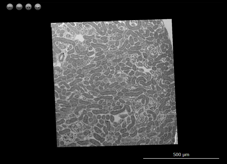
Sample : Mouse kidney
Imaging Device : JEM-1400 / Matataki Flash (2,048 x 2,048 pixels)
Image acquisition area : H 800 μm x W 760 μm with 8,640 (H 96 x W 90) images
Pixel size of the image : 5.6 nm/pixel
Total number of the pixels : 20 Gigapixel
Acceleration Voltage : 80 kV
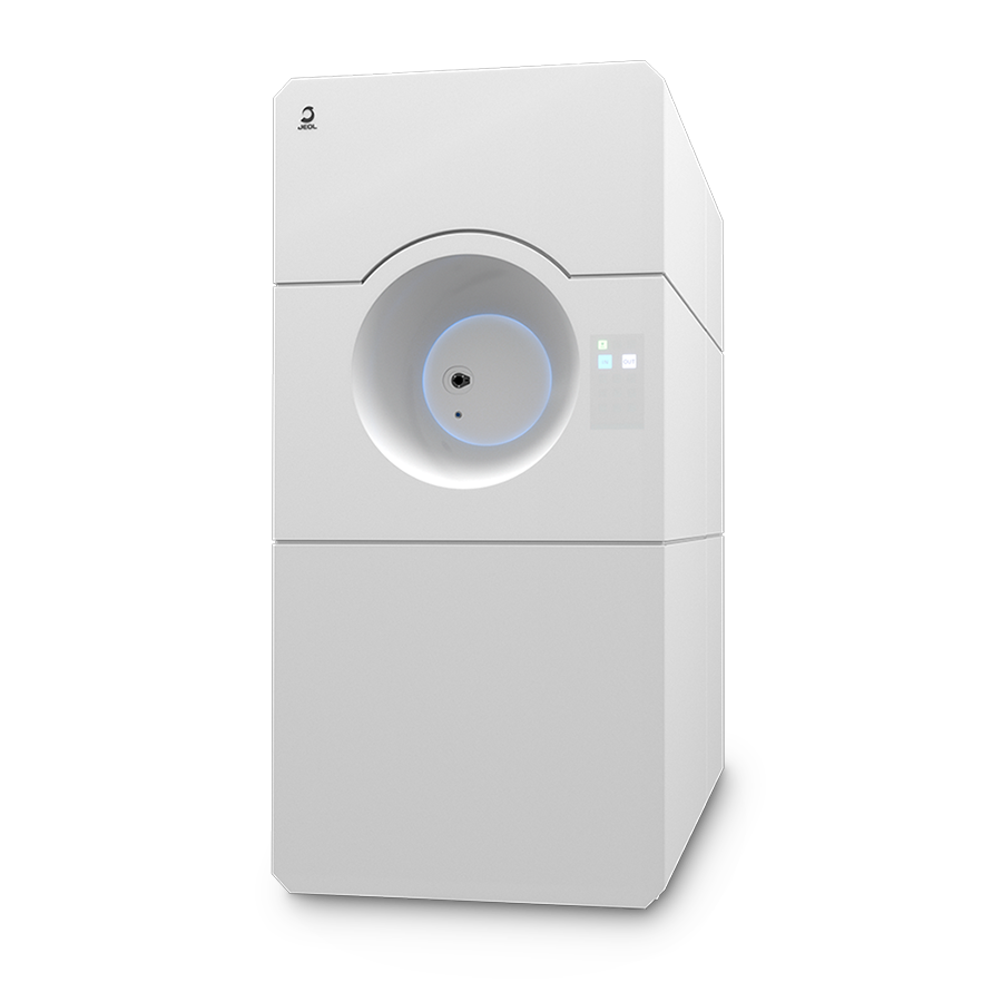
JEM-120i Electron Microscope
Transmission Electron Microscopes (TEM) with 120 kV accelerating voltage are widely used in soft material fields such as biology and polymer. We have newly developed the JEM-120i with the concept of "Compact", "Easy To Use", and "Expandable". With the new external appearance, this instrument has evolved into a useful tool that anyone can use easily, from operation to maintenance.
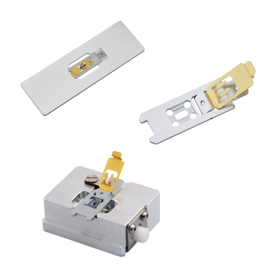
SiN Window Chip
The high-strength SiN film enables us to observe a serially large area of a millimeter in size. It is also ideal for the observation of serially sliced sections because there is no invisible area that is caused by conventional TEM grids. The dedicated retainer makes it easy to perform Correlative Light and Electron Microscopy (CLEM).
Click the button below to view more Clinical / Pathological Tests applications.