Gold particles eaten by paramecium
EM2020-02E
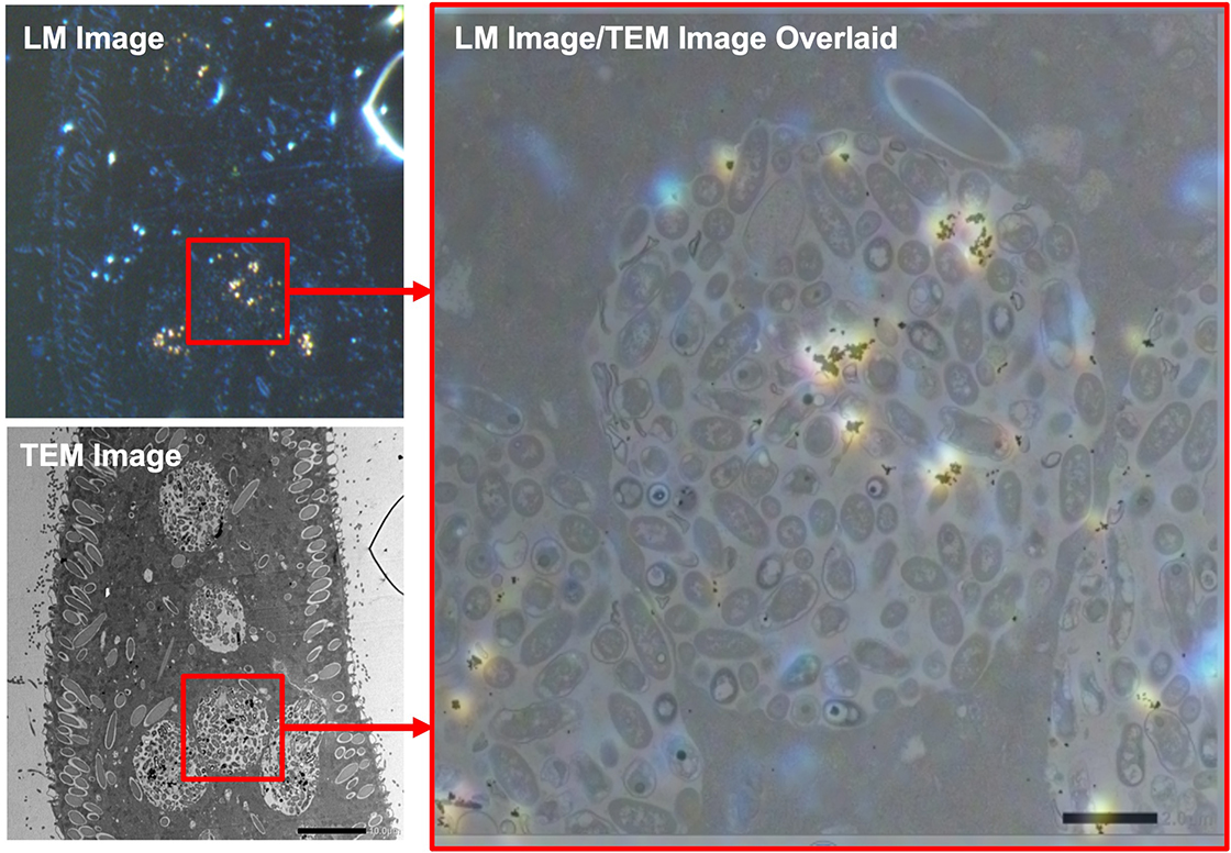
Cultured paramecium in culture medium with gold nano particles was prepared by a standard TEM protocol. Gold nano particles appear in orange by a phenomenon of surface plasmon resonance (SPR) in a dark field view with a light microscope (shown in LM image). As a result of TEM observation of the orange area that was observed by using LM, food vacuoles which had endocytosed gold particles were observed (shown in TEM image). Overlaying the LM image and the TEM image shows that localizations of gold nano particle in the TEM image were coincident with localization of the orange area in the LM (shown in the overlaid image).
Sample: Paramecium
Imaging device: Nikon Eclipse LV100DA-U, TEM: JEM-1400Flash
- Please see the PDF file for the additional information.
Another window opens when you click. 
PDF 525.0 KB
Related Products
Are you a medical professional or personnel engaged in medical care?
No
Please be reminded that these pages are not intended to provide the general public with information about the products.