Observation of TOM20 localization in HeLa cell
EM2020-03E
After making TOM20 antibody, a mitochondria marker reacted, secondary antibodies labelled with gold nano particles were made reacted.
Then, the particle diameter of a gold nano particle was sensitized to 20 nm or larger by gold sensitization. The gold particles of 20 nm or larger emit scattering light in orange due to surface plasmon resonance (SPR) when its dark field is observed with a light microscope (LM). When the same field of view is observed by TEM, mitochondria can be observed. The result shows that TOM20 are localized on the mitochondria film since gold nano particles are localized on the mitochondria film.
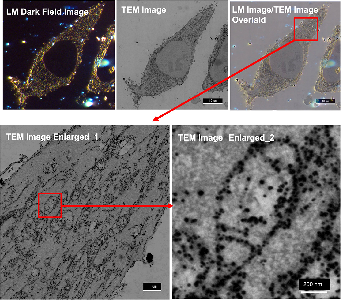
Sample HeLa cells, primary antibody: rabbit anti-TOM20 antibody, second antibody: goat anti-rabbit Fab', nanogold
Imaging device Dark field image: Nikon Eclipse LV100DA-U, 60x TEM: JEM-1400Flash
- Please see the PDF file for the additional information.
Another window opens when you click. 
PDF 582.0 KB
Related Products
Are you a medical professional or personnel engaged in medical care?
No
Please be reminded that these pages are not intended to provide the general public with information about the products.