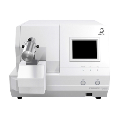
IB-10500HMS
CROSS SECTION POLISHER™
High Throughput Milling system
Features
High Throughput Milling *1
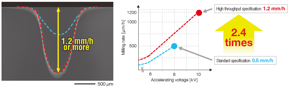
High milling rate of cross-section achieved by the new ion source:1.2mm/h or more*2
(2.4 times than the previous milling rate.)
The high throughput milling system optimizes the ion source electrodes and enables higher accelerating voltages, thus improving the ion-beam current density.
Our newly developed ion source achieves a high milling rate of cross-section of 1.2 mm/h or more (2.4 times than the previous milling rate.)
Cross-section milling rate of the new ion source
Specimen: Silicon wafer
Accelerating voltage: 10kV
Milling time: 1h
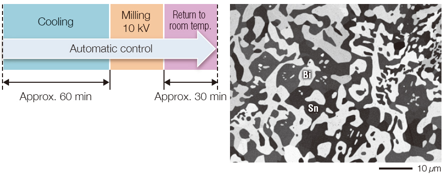
Cross-section milling of a low melting-point alloy (cooling)
Accelerating voltage: 10 kV
Milling time: 30 min
The right SEM image shows an Sn-Bi alloy with a melting point of 150°C.
A low melting-point metal can be melted due to the processing heat; therefore, cooling of the metal is required before milling. High throughput milling is applied to the heat-sensitive specimen while the specimen is kept cooled *3.
Then, a cross-section specimen with a reduced heat damage is obtained in a short time.
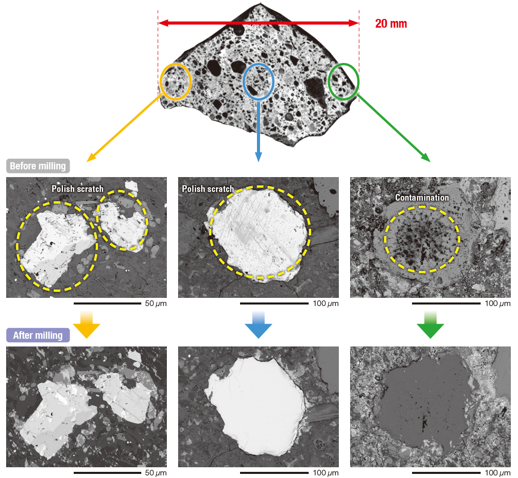
Large Area Milling *1,*4
Planar Surface Milling of Larger Area
The new high throughput milling system has enabled the irradiation of an ion beam onto a larger area of the specimen.
Planar surface milling is effective to remove scratches generated on the specimen surface or crystalline strains, which are caused by mechanical polishing.
Planar surface milling of a concrete
Accelerating voltage: 10 kV
Milling time: 20 min
Large-area planar surface milling was applied to a concrete with a width of 20mm.
After milling, polish scratches and contamination were removed, allowing for clear observation of particles of stone and cement contained in a concrete.
This function is included in IB-19530CP or IB-19520CCP which incorporates the optional high throughput specification.
Milling of 1h, Si equivalent, Edge distance: 100μm
This function is included in IB-19520CCP.
Large Specimen Rotation Holder, IB-11550LSRH, is required.
The screen images include items that are still under development, and are subject to change without notice.
Specifications
| High throughput specification ※1 | |||
|---|---|---|---|
| IB-19530CP+IB-10500HMS | IB-19520CCP+IB-10500HMS | ||
| Ion accelerating voltage | 2 to 10kV | ||
| Milling speed | 1200μm/h or more (accelerating voltage 10 kV)※2 | ||
| Specimen swing function ※3 | Auto specimen swing by ±30°,Angle setting swing | ||
| Auto milling start mode | ○ | ||
| Auto cooling milling start mode / Auto return to room temperature mode |
- | ○ | |
| Specimen stage ultimate cooling temperature | - | –120°C or less | |
| Cooling temperature settable range | - | -120 to 0°C | |
| Specimen cooling time to reach –100°C | - | Within 60min | |
| Specimen cooling retention time | - | 8h or more ※4 | |
| Air isolation function | - | - | |
| Intermittent milling mode | Ion beam irradiation time and stop time are settable (ON: 1 to 999s, OFF: 1 to 999s) | ||
| Fine milling mode | Milling conditions automatically switched | ||
| Large-area cross-section milling mode ※5 | Maximum milling width: 8mm (with optional Large Area Milling Holder IB-11730LMH) | ||
| Large-area planar surface milling mode | ○ ※6 | ||
| Maximum specimen size |
Cross-section milling |
11mm(W)×10mm(L)×2mm(T) (with standard holder for IB-19530CP) 11mm(W)×8mm(L)×3mm(T) (with standard holder for IB-19520CCP) 25mm(W)×15mm(L)×10mm(T) (with optional Large Area Milling Holder IB-11730LMH) |
|
| Planar surface milling |
40mm(diameter)×15mm(T) (with optional Large Specimen Rotation Holder IB-11550LSRH) | ||
| Specimen movements | X-axis: ±6mm, Y-axis: ±2.5mm | ||
| Operation | Touch panel, 6.5-inch display | ||
| Positioning for milling | Monitor from above the specimen stage with a camera ※7. Milling position is adjustable with an optical microscope. |
||
| Positioning camera (magnification) | Approx. ×70 (on 6.5-inch display) | ||
| Monitoring camera (magnification) | Approx. ×20 to 100 (on 6.5-inch display) *Note: When used with IB-19530CP+IB-14510MCAM ※8 or IB-19520CCP. |
||
| External monitor output | Positioning camera and Monitoring camera can be switched for displaying one on the external monitor. *Note: When used with IB-19530CP + IB-14510MCAM ※9 or IB-19520CCP + EC-10020VST ※9. |
||
| Preset function | 4 sets of milling conditions (accelerating voltage, Ar gas flow, milling time, intermittent milling) | ||
| Dimensions and weights | Basic unit | 545mm(W)×550mm(D)×420mm(H), Approx. 66kg (with IB-19530CP + IB-14510MCAM attached) 690mm(W)×720mm(D)×530mm(H), Approx. 75kg (with IB-19520CCP attached) |
|
| Rotary pump | 120mm(W)×288.5mm(D)×163mm(H), Approx. 9.3kg | ||
Installation Requirements
| Power supply | Single phase 100 to 120V AC, 50/60Hz, Allowable input voltage fluctuation: less than 10%, Rating: 15A or more |
|---|---|
| Maximum power consumption | 650VA |
| Grounding | 100 Ω or less |
| Argon gas ※10 | Dry argon, Purity: 99.9999% or more. Pressure: 0.1 to 0.2MPa (1.0 to 2.0kgf/cm2), Hose joint: ISO 7/1 Rc 1/4 |
| Room temperature | 15 to 25°C |
| Room humidity | 60% or less (no condensation) |
This is optional, which is added at the time of shipment from factory.
Milling of 1 h, Si equivalent, Edge distance 100 μm
Patent No. (Japan): 4557130
As the set temperature is higher, the cooling retention time is longer.
This mode can be used in combination with the cooling function of IB-19520CCP.
When used with IB-11550LSRH.
Patent No. (Japan): 4208658
With IB-14510MCAM attached, the specimen can be monitored in real time.
The status of the specimen can be observed while milling is in progress.
The external monitor must be prepared by the customer.With EC-10020VST attached, the camera image can be displayed on the external monitor.
The external monitor must be prepared by the customer.The argon gas, gas cylinders and regulator must be prepared by the customer.
The specifications and appearance of the instrument are subject to change without notice.
Catalogue Download
IB-10500HMS CROSS SECTION POLISHER™ High Throughput Milling system
Related Products
Related Products
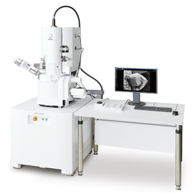
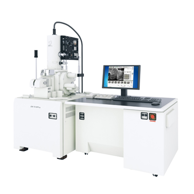
JSM-IT800 Schottky Field Emission Scanning Electron Microscope
The JSM-IT800 incorporates our "In-lens Schottky Plus field emission electron gun" for high resolution imaging to fast elemental mapping, and an innovative electron optical control system "Neo Engine", as well as a system of seamless GUI "SEM Center" for fast elemental mapping with a fully embedded JEOL energy dispersive X-ray spectrometer (EDS), as a common platform.
The JSM-IT800 allows for the replacement of the objective lens of the SEM as a module, offering different versions to satisfy various users requirements. With the JSM-IT800, five versions are available with different objective lenses: a hybrid lens version (HL), which is a general-purpose FE-SEM; a super hybrid lens version (SHL/SHLs, two versions with different functions), which enables higher resolution observation and analysis; and the newly-developed semi-in-lens version (i/is, two versions with different functions), which is suited for the observation of semiconductor devices.
Furthermore, the JSM-IT800 can also be equipped with a new Scintillator Backscattered Electron Detector (SBED) and a Versatile Backscattered Electron Detector (VBED). The SBED enables the acquisition of images with high responsiveness and produces sharp material contrast even at a low accelerating voltage, while the VBED can help obtain images of 3D, topography and material contrasts. Thus, the JSM-IT800 can help users to obtain information that was not obtainable and to solve problems in measurement.
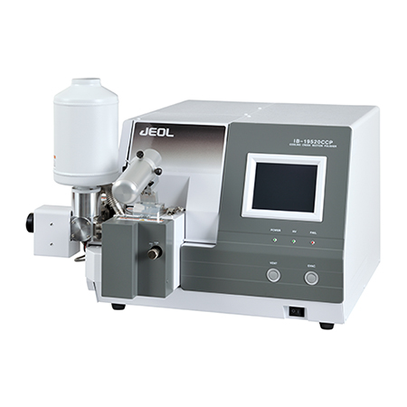
IB-19520CCP CROSS SECTION POLISHER™
Thermal damage can be reduced by cooling the specimen with liquid nitrogen during processing.Designed to suppress the consumption of liquid nitrogen, allowing long cooling periods.<br>Rapid cooling of the specimen while immersed in liquid nitrogen. Return to room temperature. Designed to allow parts to be detached.<br>Incorporates a mechanism to allow the process from polishing to observation to be performed without exposing the sample to the air.
IB-19530CP CROSS SECTION POLISHER™
The IB-19530CP features an innovatively designed, multi-purpose stage to fulfill increasingly diversified market needs and realize multi-functionality by different kinds of functional holders. The multi-purpose stage combined with specialized functional holders allows the user to perform various functions such as planar surface milling and polishing, sputter coating as well as more traditional cross-section ion milling.
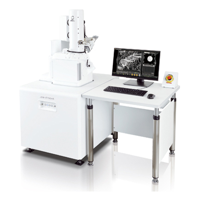
JSM-IT700HR InTouchScope™ Scanning Electron Microscope
SEM- Essential in Daily Lab Operation JSM-IT700HR Makes it Easy.
Nano-scaled materials are driving the current technological breakthroughs and their observation and analysis is facilitated by a new and innovative SEM, JSM-IT700HR.
Its new electron gun with spatial resolution of 1 nm and the largest probe current of 300 nA, combined with an exceptionally userfriendly software interface significantly simplifies observation and analysis in SEM.
The compact instrument design also features a large specimen chamber with multiple accessory ports as well as EDS integration.
JSM-IT700HR Advanced SEM, Powerful and Simple to Use.
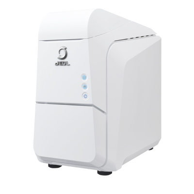
JCM-7000 NeoScope™ Benchtop SEM
Benchtop scanning electron microscopes are used in a wide range of fields, such as electrical, electronics, automobiles, machinery, chemical, and pharmaceutical industries. In addition, SEM applications are expanding to not only cover research and development, but also address quality control and product inspection at manufacturing sites. With this, demands for further improved work efficiency, much faster and easier operation, and a higher degree of analytical and measurement capabilities, are increasing.
The JCM-7000 Benchtop Scanning Electron Microscope is designed based on a key concept of "Easy-to-use SEM with seamless navigation and live analysis". The JCM-7000 incorporates three innovative functions; "Zeromag" for smooth transition from optical to SEM imaging, "Live Analysis" for finding constituent elements for an image observation area, and "Live 3D" for displaying a reconstructed live 3D image during SEM observation.
When you place the JCM-7000 next to an optical microscope, further-faster and more-detailed foreign material analysis and quality control can be made.
More Info
Are you a medical professional or personnel engaged in medical care?
No
Please be reminded that these pages are not intended to provide the general public with information about the products.