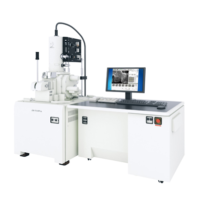
【DISCONTINUED】JSM-7610FPlus Schottky Field Emission Scanning Electron Microscope
DISCONTINUED
This product is no longer available.
If you would like to know the latest information about your preferred product or to find out more about alternatives, please click on the link below. We hope you will continue to use our products.
Features
Features
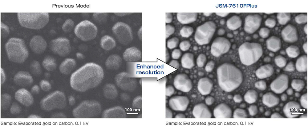
The highly-acclaimed optical system of the JSM-7610F has been updated, achieving even better resolution (15 kV 0.8 nm, 1 kV 1.0nm), and is now available as the JSM-7610FPlus.
The Semi-in lens type objective lens and High Power Optics of the irradiation system deliver high-spatial resolution observation and stable analysis capability.
In addition, the JSM-7610FPlus can be equipped to satisfy a variety of user needs, including observation at low accelerating voltages with GENTLEBEAM™ mode, and signal selection using r-filter.
Semi-in Lens Objective Lens
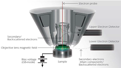
Semi-in lens objective lens design allows ultra-high resolution observation with in-lens detector.
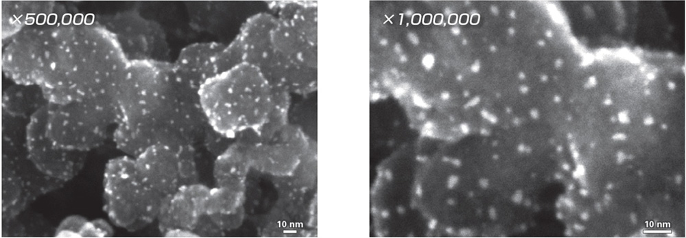
Sample:Platinum Catalyst Nano Particles on Carbon 15kV
High Power Optics
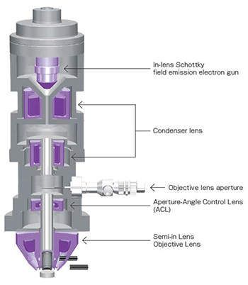
A unique electron optical system allows a variety of analyses and observation at high magnification. The in-lens Schottky field emission electron gun, which can deliver a probe current 10 times that of the conventional Schottky field emission electron gun (FEG), along with the aperture-angle control lens (ACL) that can maintain a small probe diameter with the appropriate convergence angle even with the increased probe current, makes it possible to use probe currents of 200 nA or more. The high-power optics provides all the performance you need to conduct everything from high-magnification viewing to EDS and EBSD analyses.
In-lens Schottky Electron Gun
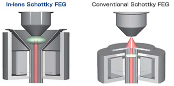
The in-lens Schottky field emission electron gun combines the electron gun with a low-aberration condenser lens to enable efficient collection of the electrons generated from the electron gun.
The Aperture-Angle Control Lens (ACL)
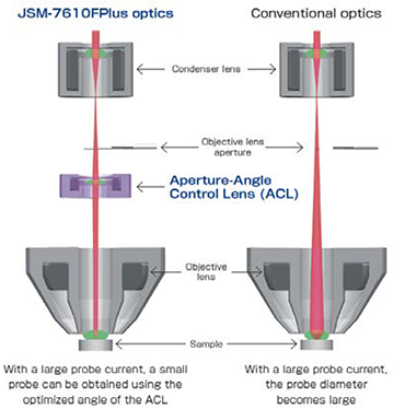
The Aperture-Angle Control Lens (ACL) is positioned above the objective lens to perform automatic optimization of the objective lens aperture angle across the entire probe current range. This makes it possible to obtain a smaller probe diameter than is possible with conventional systems, even when the probe current is large.
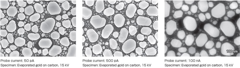
Small Probe Diameter, Even With Large Probe Current
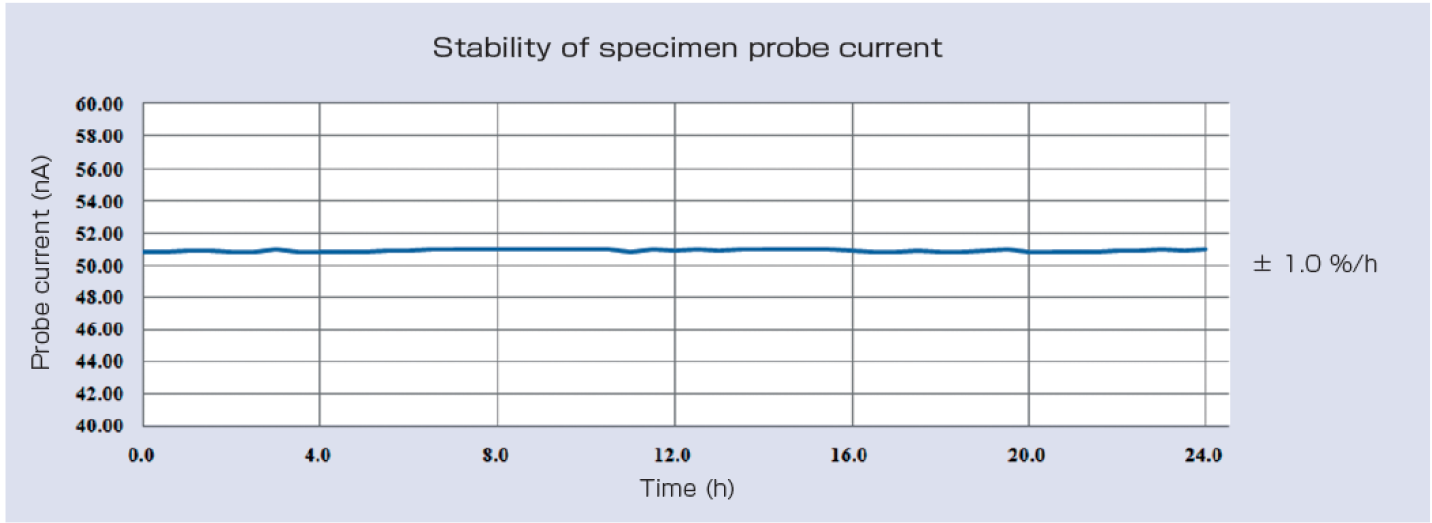
Highly Stable Probe Current for extended analysis times
The in-lends Schottky field emission electron gun delivers a stable probe current.
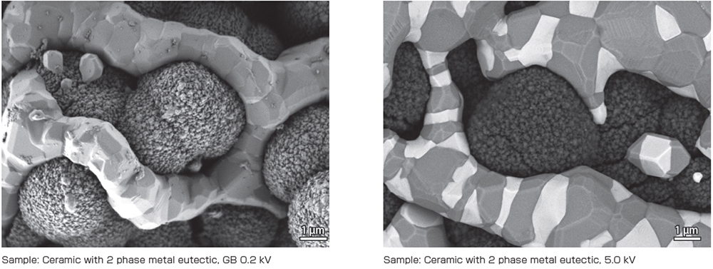
GENTLEBEAM™mode
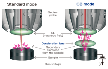
In GENTLEBEAM™ mode (GB mode) , a voltage is applied to the specimen to reduce the landing voltage of the electrons just before they strike the specimen, enabling high-resolution observation with accelerating voltages as low as 100 V.
Since the scattering region of the electron beam within the specimen is small, it is easy to observe the micro structures on the surface, and the influence on specimens that are susceptible to heat damage can be reduced. Non-conductive specimens can be easily observed with reduced charging.
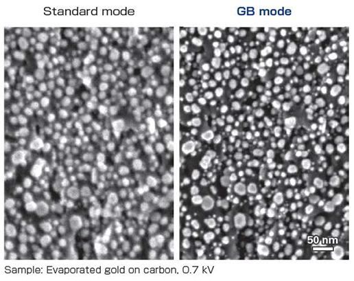
The GB mode 1 kV resolution with the JSM-7610FPlus is 1.0 nm.
Effect of GB Mode
GB mode improves resolution at low accelerating voltage.
Improved Resolution at Low Accelerating Voltage
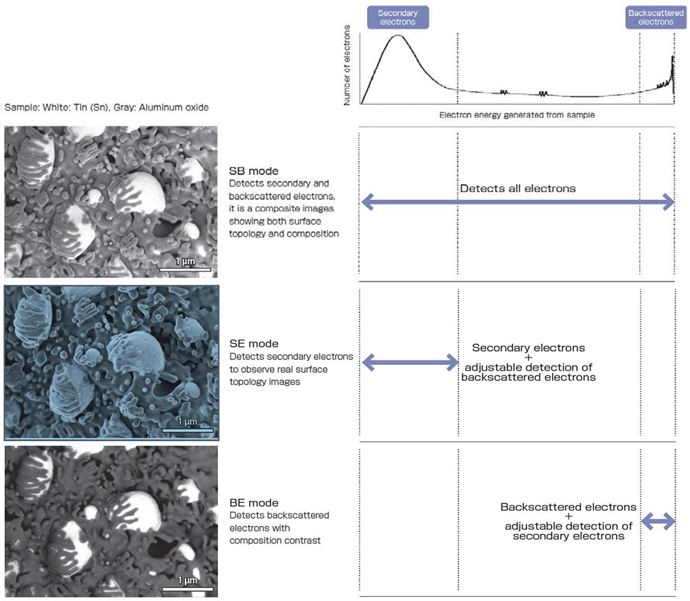
r-filter
Next- generation r-filter
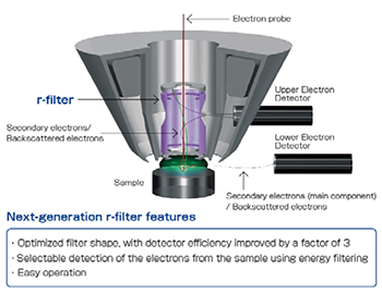
The next-generation r-filter is a unique energy filter that combines a secondary electron control electrode, a backscattered electron control electrode and a filter electrode. When the specimen surface is irradiated by the electron beam, electron with various energies are emitted from the surface. The new r-filter makes it possible to selectively detect the secondary electrons and backscattered electrons from the specimen while the electron beam is held at the center of the lens using a combination of multiple electrostatic fields.
In comparison to the r-filter of the previous model, the next-generation r-filter has a 3 fold increase in signal.
Signal Detection With The New r-filter
LABE Detector (Option)

The LABE (Low Angle Backscatter Electron) detector is capable of detecting very low energy and very low angle backscattered electrons that were previously not able to be detected.
Detailed topological informations of the specimen surface can be obtained at extremely low accelerating voltages, and compositional information for the specimen can be observed with good resolution at high accelerating voltages.
LABE Detector: Low Angle Backscattered Electron Images
Thin-film crystal grain channeling contrast can be observed using backscattered electrons with an extremely low accelerating voltage.
Extensibility
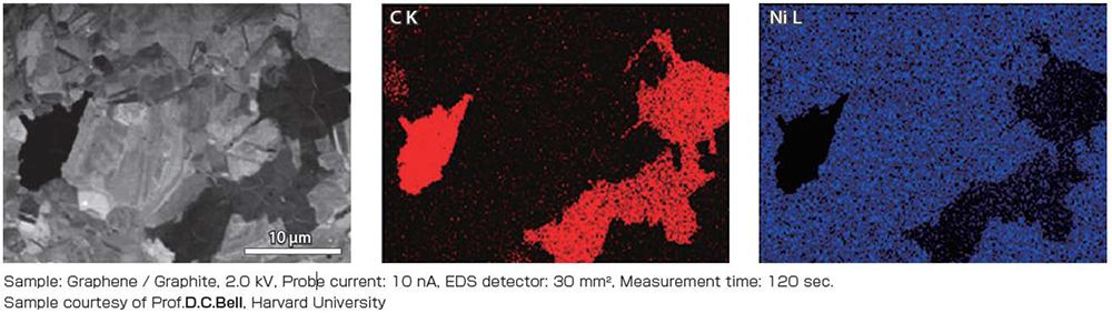
Energy Dispersive X-ray Spectrometer (EDS)
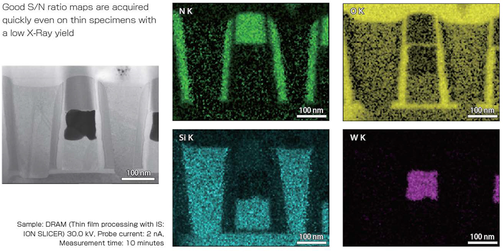
The High Power Optics make it possible to effectively take advantage of the features of the EDS (SDD: Silicon Drift Detector) detector that is difficult to saturate even with large probe currents. By using a low accelerating voltage and a large probe current, good quality mappings can be obtained in a very short period of time. The images below show the analysis of an extremely thin graphene and graphite layer on a Ni substrate obtained in 2 minutes.
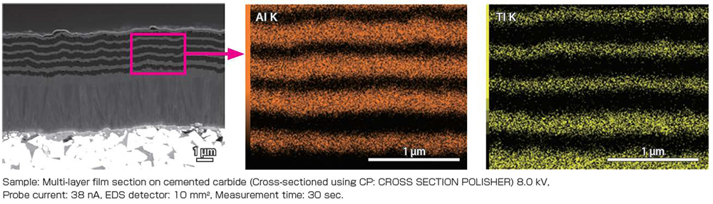
Even using basic type SSD 10 mm2 detector it is possible to analyze the cross section of a multi-layer film of about 100 nm in just 30 seconds.
Wavelength Dispersive X-ray Spectrometer (WDS)
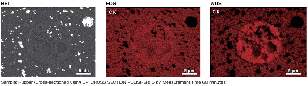
Because the High Power Optics provide a large probe current with a small probe diameter, it is possible to take full advantage of the features of WDS. Using WDS enables confirmation of trace concentration differences or elemental overlaps in a specimen, which cannot be identified with EDS.
Cathodoluminescence Detector (CL)
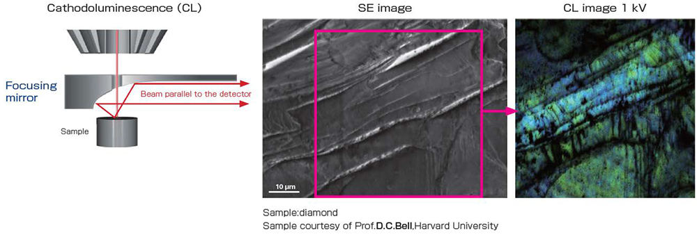
CL is a phenomenon of visible light being generated when a specimen is exposed to an electron beam. The light that is generated from the specimen is collected using a focusing mirror and detected. The images below are a secondary electron image and a CL image obtained with a diffraction lattice of diamond at 1 kV. Observing the CL image with a low acceleration voltage reveals defects in the diamond surface with good resolutuion.
Specimen Chamber Optimized for Analysis
The specimen chamber is designed to permit installation of the various detectors in an optimal layout, including the secondary electron detector, backscattered electron detector, EDS, EBSD, WDS, STEM, and cathodoluminescence detector.
The secondary electron detector, EDS and EBSD are positioned to enable them to be seen at the same time ona a tilted specimen, with the EBSD port perpendicular to the eucentric tilt on the stage.
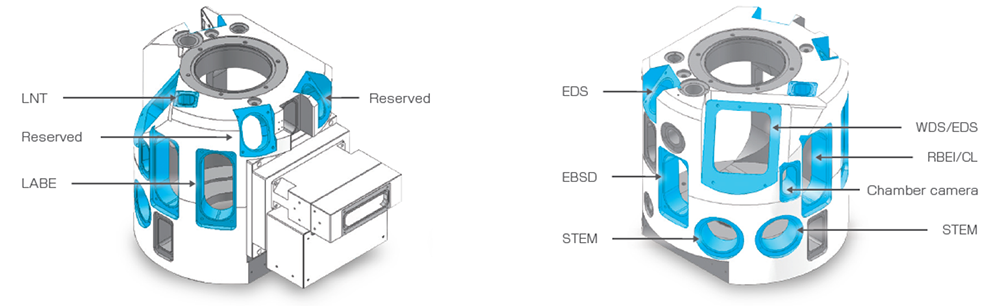
A variety of accessories can be installed simultaneously
Specifications
| Secondary electron image resolution |
0.8 nm(Accelerating voltage 15 kV) 1.0 nm(Accelerating voltage 1 kV GB mode) 0.8 nm(Accelerating voltage 1 kV GBSH mode)※1 During analysis 3.0 nm (Accelerating voltage 15 kV, WD 8 mm, Probe current 5 nA) |
||
|---|---|---|---|
| Magnification |
Direct magnification: x25 to 1,000,000(120 x 90mm) Display magnification: x75 to 3,000,000(1,280 x 960 pixels) |
||
| Accelerating voltage | 0.1 to 30 kV | ||
| Probe current | A few pA to ≥ 200 nA | ||
| Electron Gun | In-lens Schottky field emission electron gun | ||
| Lens system |
Condenser lens (CL) Aperture-angle control lens (ACL) Semi-in lens objective lens (OL) |
||
| Specimen stage | Fully eucentric goniometer stage | ||
| Specimen movement |
Specimen stage Standard |
Optional | Optional |
|
type I A2 X : 70 mm Y : 50 mm Z : 1.0 to 40 mm Tilt: -5 to +70° Rotation: 360° |
type II X : 110 mm Y : 80 mm Z : 1.0 to 40 mm Tilt: -5 to +70° Rotation: 360° |
type III X : 140 mm Y : 80 mm Z : 1.0 to 40 mm Tilt: -5 to +70° Rotation: 360° |
|
| Specimen holders | 12.5 mm diameter × 10 mm thick, 32 mm diameter × 20 mm thick | ||
| Specimen exchange | One-action exchange mechanism | ||
| Electron detector system | Upper detector, r-filter, Built-in, Lower detector | ||
| Automatic Functions | Focus, Stigmator, Brightness, Contrast | ||
| Image observation LCD | Screen size: 23-inch wide Maximum resolution: 1,280 × 1,024 pixels |
||
| SEM Control System | PC: IBM PC/AT compatible computer OS: Windows® 7 Professional※2 |
||
| Scan and display modes |
Full-frame scan Real magnification Selected- area scan Two-image display (with different magnifications, different image modes) Two-image wide display Four-image display (four-signal live display) Addition image (4 images + addition image) Scale |
||
| Evacuation System |
Gun chamber, first and second intermediate chambers: Ultra high-vacuum dry evacuation system using ion pumps Specimen chamber: Dry evacuation system using a turbo-molecular pump (TMP) |
||
| Ultimate pressure |
Gun chamber: Order of 10-7 Pa (for standard configuration) Specimen chamber: Order of 10-4 Pa (for standard configuration) |
||
Optional
Microsoft Windows is registered trademark of Microsoft Corporation in United States and other countries
Catalogue Download
JSM-7610FPlus Schottky Field Emission Scanning Electron Microscope
Application
Application JSM-7610FPlus
More Info
Are you a medical professional or personnel engaged in medical care?
No
Please be reminded that these pages are not intended to provide the general public with information about the products.