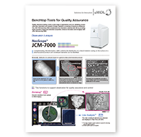
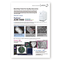
Benchtop scanning electron microscopes (also known as a tabletop SEM or desktop SEM) are used in a wide range of fields, such as electrical, electronics, automobiles, machinery, chemical, and pharmaceutical industries. In addition, SEM applications are expanding to not only cover research and development, but also address quality control and product inspection at manufacturing sites. With this, demands for further improved work efficiency, much faster and easier operation, and a higher degree of analytical and measurement capabilities, are increasing.
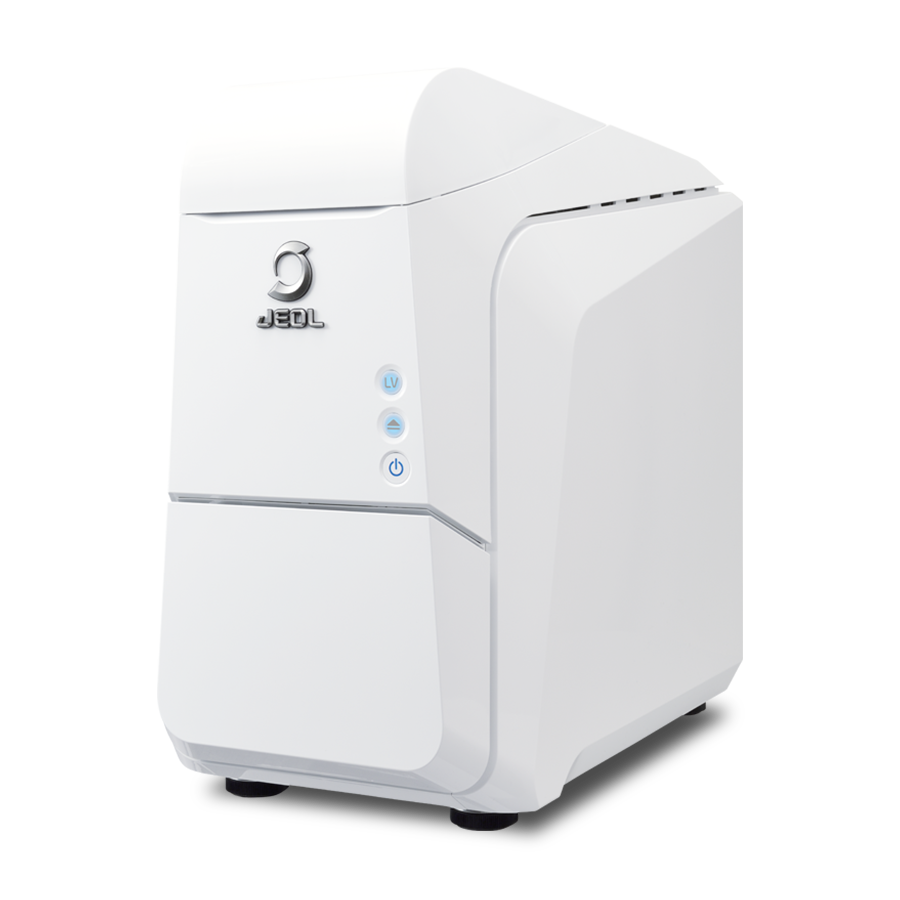
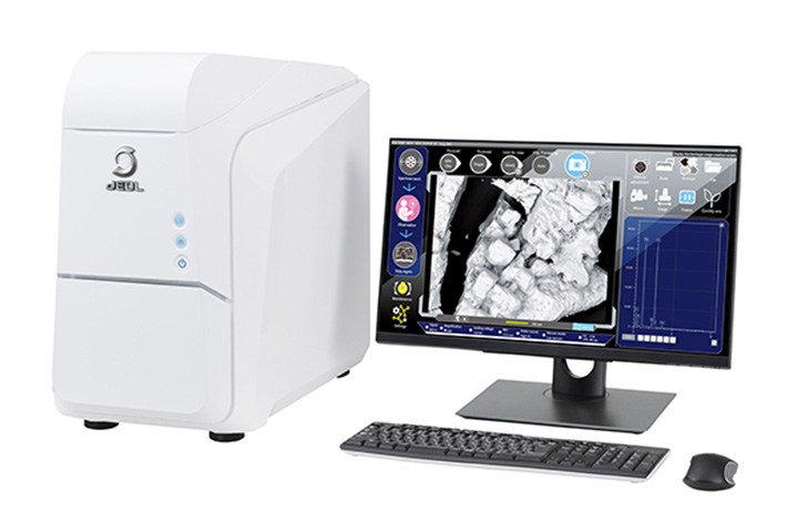
The JCM-7000 Benchtop Scanning Electron Microscope is designed based on a key concept of "Easy-to-use SEM with seamless navigation and live analysis". The JCM-7000 incorporates three innovative functions; "Zeromag" for smooth transition from optical to SEM imaging, "Live Analysis" for finding constituent elements for an image observation area, and "Live 3D" for displaying a reconstructed live 3D image during SEM observation.
When you place the JCM-7000 next to an optical microscope, further-faster and more-detailed foreign material analysis and quality control can be made.
Features
Download
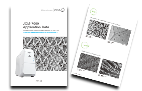
Speedy observation and analysis with no specimen treatment using the benchtop SEM JCM-7000!
-Introduction to the effective use of Low-Vacuum(LV) mode-
Why don't you try an SEM observation of insulating specimens that do not conduct electricity as they are, without any pre-treatment?
We will introduce examples of effective use of the JCM-7000 in a low vacuum (LV) mode for polymeric materials, industrial materials, minerals, foods, and living organisms/plants.
Conventional SEM
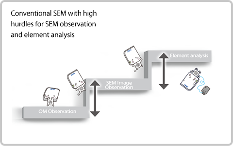
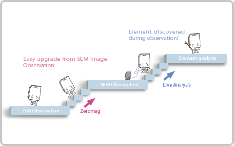
In conventional SEM operation, SEM imaging and elemental analysis were separated (not seamless).
JCM-7000
With "Zeromag", the JCM-7000 enables seamless operation from optical to SEM imaging. Also with "Live Analysis", elemental analysis by EDS can be made during SEM image observation.
Comparison with OM
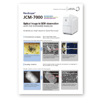
Accelerate your insight beyond an Optical Microscope
Contaminant analysis
Easy to detect foreign material
Easy to identify elemental composition
【Example】 Analysis of black foreign material adhered to surface of food product
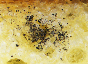
OM image
In OM image, it is difficult to see the distribution of the whilte lubricant on the white granule(drug) surface and quality of its adhesion.

SEM Image (Backscattered electron compositional image)
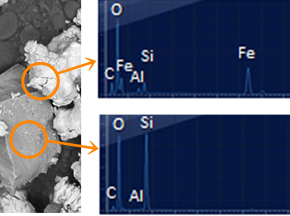
SEM image from the same field of view (FOV) shows particles with different contrast indicating different compositions.
SEM Image (Backscattered electron compositional image) and elemental analysis result
Enlarging the area of interest accesses instant live EDS analysis with main elements identified.
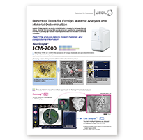
Quality control
Observe detailed surface structures with high resolution and large depth of field not possible with OM imaging.
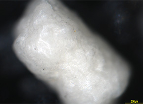
【Example】 Distribution observation of lubricant on the surface of granule(drug)
OM image
In OM image, it is difficult to see the distribution of the lubricant on the granule surface and quality of its adhesion.
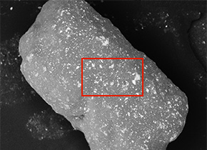
SEM Image(Backscattered electron compositional image)
The superior depth of focus provided with SEM imaging over OM imaging along with the compositional contrast provided with the backscattered electron detector clearly shows the distribution of the lubricant on the surface of the granule.
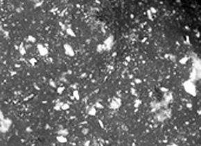
SEM Image (Backscattered electron compositional image) and elemental analysis
Condition of the lubricant's adhesion can be observed with higher magnification.
Features and Applications
Functions that enable anyone to perform SEM/EDS operations
Zeromag & Low-Vacuum mode
Zeromag
Zoom the optical image to automatically switch to a SEM image!
Low-Vacuum mode
Viewing is simple with no pre-treatment needed for easily-charged samples.
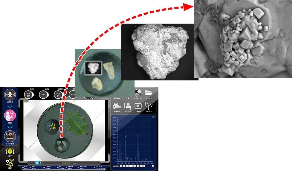
Specimen: Salt
Click the "replay" button in the box above, and the movie will start (for 60 seconds)
Live! Live! Live!
Live Analysis
eliminates the need to consider SEM observation and EDS analysis as separate operations. When Live Map is selected, you can confirm the distribution of elements in the observed area in real-time.
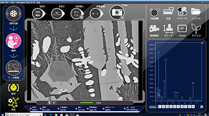
Screening while performing observation with Live Analysis
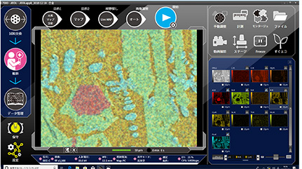
Quickly check the element distribution with Live Map
Live 3D
The new high-sensitivity 4-segmented backscattered electron detector enables 2-pane viewing of a SEM image and a 3D image using Live 3D function. In addition to instantaneous shape determination for samples with complex topographies, depth information can also be acquired.
SMILE VIEW™ Lab
All data can be managed from SMILE VIEW™ Lab Data management screen.
Review and re-analysis of previously-acquired data, and creation of reports can be performed easily.
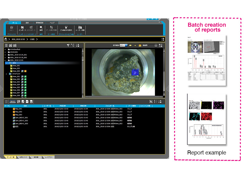
Click the "replay" button in the box above, and the movie will start (2.5 min.)
This movie introduces you to the features and functions of Benchtop SEM JCM-7000 NeoScope™.
Function explanation by movies
The following movie introduces you to the functions of the Benchtop SEM JCM-7000 NeoScope™
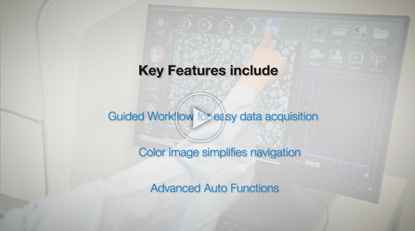
Three features for easy operation
The JCM-7000 is characterized by its ease of use. Here are three features that even beginners will love.1.Guided Workflow for easy data acquisition2.Color image simplifies navigation3.Advanced Auto Functions
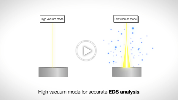
Suitable for observation and analysis of a variety of specimens – High vacuum mode provides higher quality measurements! –
The JCM-7000 is equipped with a high vacuum mode for observation and analysis of conductive specimens and low vacuum mode for observation and analysis of conductive specimens with no specimen treatment.Therefore, the JCM-7000 can be used for SEM observation and EDS analysis of a diverse range of specimens.
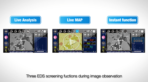
Leave both observation and analysis to us
JEOL develops and manufactures both SEM and EDS in-house.Both SEM observation and EDS analysis can be operated at an optimal position and within the same user interface.The movie introduces you to the three EDS screening functions during image observation.
Download
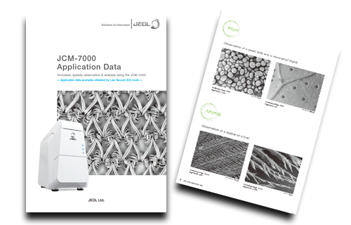
Speedy observation and analysis with no specimen treatment using the benchtop SEM JCM-7000!
-Introduction to the effective use of Low-Vacuum(LV) mode-
Why don't you try an SEM observation of insulating specimens that do not conduct electricity as they are, without any pre-treatment?
We will introduce examples of effective use of the JCM-7000 in a low vacuum (LV) mode for polymeric materials, industrial materials, minerals, foods, and living organisms/plants.
Application
Application JCM-7000
Cross Sections of Noodles Observed at Various Cooling Temperatures
Observation Examples of Processed Foods
More Info
Are you a medical professional or personnel engaged in medical care?
No
Please be reminded that these pages are not intended to provide the general public with information about the products.

