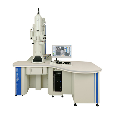
【DISCONTINUED】JEM-1400Plus Electron Microscope
DISCONTINUED
This product is no longer available.
If you would like to know the latest information about your preferred product or to find out more about alternatives, please click on the link below. We hope you will continue to use our products.
The JEM-1400Plus is a transmission electron microscope (TEM) developed for application in a wide range of disciplines, from biology to materials researches, such as biological sections, polymers, nanomaterials and …. New environment is optimized for ease-of -use TEM operations with followings.
Features
- New operation panel and LCD screen with touch panel offering simple and ease-of-use operations.
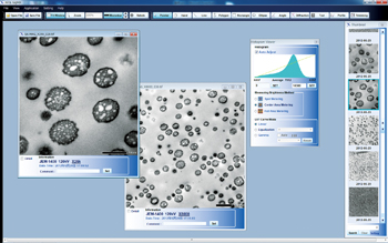
- High-precision camera (8 M pixels) fully-integrated with the PC controlled TEM operation system.
- Off-line viewer software [SightX] enabling to review and edit acquired images on a user’s PC in office.
- A wealth of automated functions, including auto-focus, auto-exposure, and auto-montage.
- New image forming lens system, delivering a magnification of x10, and enabling to acquire an entire mesh image.
One camera covers all magnifications, from searching a target to recording a final image.
With the JEM-1400Plus, images from the ultra LOWMAG mode (min. mag. ×10) to the MAG mode (max. mag. ×1.2 M) can be acquired with only one camera, resulting seamless observation with no switching of cameras or shifting one’s gaze to a fluorescent screen. Using the auto montage function (provided as standard) makes it easy to acquire high-precision images of a wide field of view. 8M pixel camera (high-resolution camera) and a 1 M pixel cameras are selectable depending on user’s purposes.
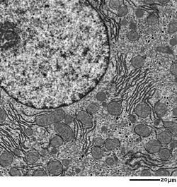
CCD camera image (8 M pixels)
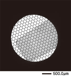
10× CCD camera image
Sample: Rat hepatic cells
Sample courtesy of Dr. Shogo Muranaka, Hamamatsu University School of Medicine
Point&Shoot function
With this function, user allows to move a field of view to target position pointed by clicking on a previously-acquired image. The Point&Shoot function allows users to view a target image without changing optical conditions such as focus or magnification.
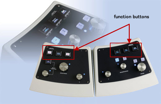
Intelligent Panel
An advanced-function, simply-designed operation panel was developed. The rich and various patterns on the color display of the organic EL enable to display a function of buttons with easy-to-see and user-friendly accessibility. About 50 kinds of button patterns are provided and the functions of the buttons are customizable on the user’s selection.
Other functions
Following software functions are prepared for user’s special purposes.
- Length measurement tools
- Image file thumbnail display
- Integration with drift correction
Specifications
| Configuration*1 | High Contrast (HC) | STEM (ST) | |
|---|---|---|---|
| Resolution (nm) | TEM | TEM | STEM*2 |
| Particle image | 0.38 | 0.38 | - |
| Lattice image | 0.2 | 0.2 | - |
| STEM bright-field image (Edge to edge) |
- | - | 2.0 |
| Accelerating voltage Minimum variable |
40、60、80、100、120kV 33V (U*with correction) |
40、60、80、100、120kV 33V (U*with correction) |
|
| Magnification | TEM mode | TEM mode | STEM mode |
| MAG mode | ×200~1,200,000 | ×200~1,200,000 | ×5,000~2,000,000 |
| LOW MAG mode | ×10~1,000 | ×10~1,000 | ×120~4,000 |
| SA MAG mode | ×2,000~300,000 | ×2,000~300,000 | - |
| Specimen tilt angle | |||
| Tilt-X | ±70°(High-tilt specimen holder) / ±25° (Standard holder) | ||
| Tilt-Y | ±9° (Using the STH holder) | ||
| Travel range | 2 mm | ||
*1 Select either configuration when the system is ordered
*2 Requires the optional scanning image observation device
Application
Application JEM-1400Plus
Ultrawide transmission electron microscopy image of a mouse kidney
Comparison of 3D Imaging Methods in Electron Microscopy for Biomaterials
Solid-State NMR Meets Electron Diffraction
Related Products
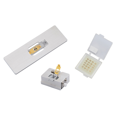
SiN Window Chip
The high-strength SiN film enables us to observe a serially large area of a millimeter in size. It is also ideal for observation of serially sliced sections because there is no invisible area that is caused by conventional TEM grids. The dedicated retainer makes it easy to perform Correlative Light and Electron Microscopy (CLEM).
More Info
Are you a medical professional or personnel engaged in medical care?
No
Please be reminded that these pages are not intended to provide the general public with information about the products.