YAG backscattered electron detector, YAG BSE detector, YAG BED
YAG backscattered electron detector, YAG BSE detector, YAG BED
A backscattered electron (BSE) detector using a crystal of Yttrium Aluminum Garnet (Y3Al5O12), abbreviated to YAG, with cerium (Ce) added as a fluorescent active material. (Here, this YAG is termed “Ce: YAG”.)
The most widely used BSE detector is the semiconductor detector. However, since the semiconductor detector has a long response time (slow response speed), a specimen image is blurred at a fast scanning speed. The YAG BSE detector is used to avoid this phenomenon.
Fig. 1 schematically shows the YAG BSE detector. Backscattered electrons, which are generated at illuminating a focused electron beam onto a specimen, travel toward a “Ce: YAG” placed just above the specimen. Then, the generated light (fluorescence light) reaches a photo-multiplier tube via a light guide. The light is converted into electrons at the photocathode and the electrons are further multiplied. The multiplied electrons reach the anode and become an electrical signal. The electrical signal is further amplified. Since “Ce: YAG” has a short attenuation time (about 70 ns) of fluorescence, the response time of the YAG BSE detector is more than one order of magnitude shorter than that of the semiconductor BSE detector.
If the response time is long, as in the case of the semiconductor BSE detector, the SEM image may be blurred as if the specimen has moved sideways when observed at a fast scanning speed (seen in the lower left image of Fig. 2). As the YAG BSE detector has a short response time, the image can be observed without blurring even at fast scanning speeds. Therefore, the image can be seen clearly even when the specimen stage is moved quickly and the desired observation position can be found without time lag. Furthermore, in the case of Array tomography, the YAG BSE detector can shorten the image acquisition time to take many images over a wide area of a specimen.
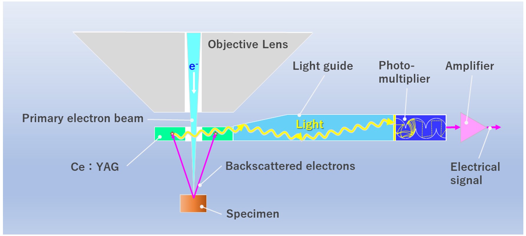
Fig. 1 Schematic of YAG BSE detector
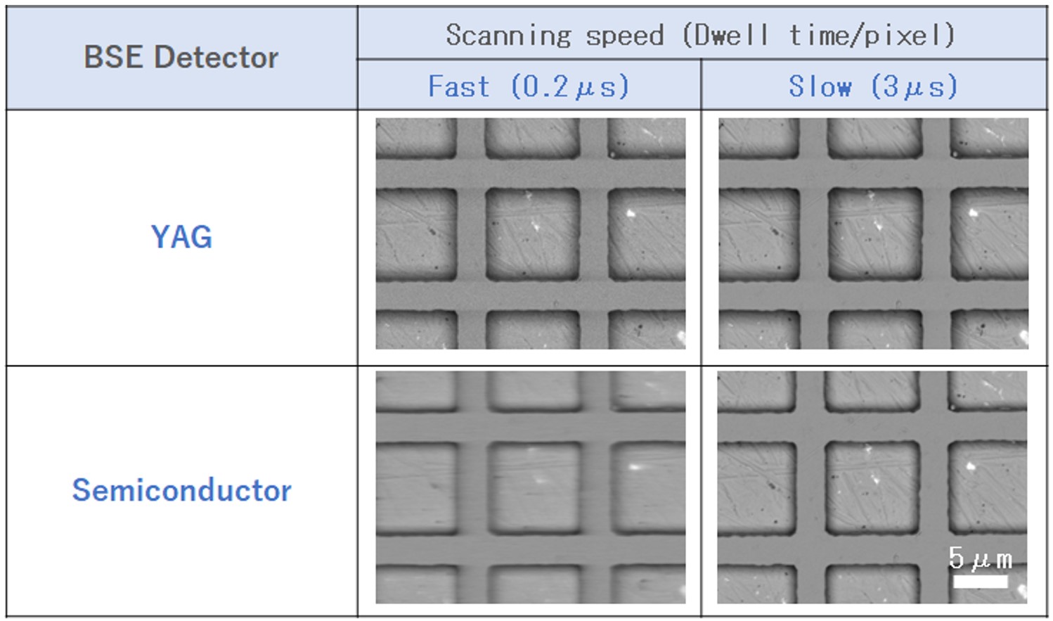
Fig. 2 BSE images acquired in a fast scanning speed (left) and a slow scanning speed (right). Blurred image (left down) is noted, as if the specimen moved sideways at a high speed scanning.
Related Term(s)
Term(s) with "YAG backscattered electron detector, YAG BSE detector, YAG BED" in the description
Are you a medical professional or personnel engaged in medical care?
No
Please be reminded that these pages are not intended to provide the general public with information about the products.


