compositional image in BE mode, compositional image in BSE mode, BSE compositional image
compositional image in BE mode, compositional image in BSE mode, BSE compositional image
"BSE compositional image" means a backscattered electron image which exhibits the difference in the average atomic number (compositional difference) in a specimen.
This image reveals the compositional distribution and the size of a mono-compositional area. In addition, the BSE compositional image is effectively used to specimen observation in advance of elemental analysis using an energy dispersive X-ray spectrometer (EDS) or a wavelength dispersive X-ray spectrometer (WDS) because the image provides an overall composition of the specimen.
For detecting backscattered electrons, an annular semiconductor device (detector) placed symmetrically against the incident (primary) electron beam is used. The detector which is divided into a pair of two segments or a pair of four segments is also available.
When the two-segmented detector is used, the BSE compositional image is obtained by adding the output signals from both pair of segments.
To the contrary, when subtracting the output signals between each pair of segments by the use of this detector, the contrast due to compositional difference is canceled out and instead, the image formed by specimen topography is obtained.
Figures below show a BSE compositional image (left) and a BSE topographic image (right) of a diamond grindstone.
In the left compositional image, diamond particles appear dark (black) because the backscattered electron emission coefficient is small for diamond. To the contrary, the regions where atoms heavier than carbon exist, are seen to be bright (white). Since the sum of the addition of two output signals from the two detectors is collected, the topographic effect in the specimen is canceled out.
In the right topographic image, the difference of the output signals between the two detectors is collected, and thus the compositional contrast disappears and only the topographic contrast of the specimen is observed.
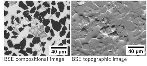
Fig. Comparison of BSE compositional image and BSE topographic image of a diamond grindstone, taken at an accelerating voltage of 10 kV
In the left compositional image, diamond particles are seen to be dark (black) because of a small backscattered electron emission coefficient of diamond. In the right topographic image, the compositional contrast disappears and only the topography of the specimen is observed.
Related Term(s)
Term(s) with "compositional image in BE mode, compositional image in BSE mode, BSE compositional image" in the description
Are you a medical professional or personnel engaged in medical care?
No
Please be reminded that these pages are not intended to provide the general public with information about the products.


