


characteristic X-rays
characteristic X-rays
Characteristic X-rays are the X-rays that are emitted when an inner-shell electron is ejected from an atom by an incident electron to create a hole, and an electron at a higher energy level in the atom transitions to fill the hole, releasing the excess energy as electromagnetic radiation. Since the energy of the characteristic X-rays has a specific value for each element, SEM can perform elemental analysis by Energy Dispersive X-ray Spectroscopy (EDS) utilizing characteristic X-rays.
Generation region of Characteristic X-rays
When incident electrons irradiate a specimen, the electrons penetrate into a certain depth while losing their energy through collisions with the atoms in the specimen. In this process, the inner-shell electrons are ejected, and characteristic X-rays are generated. The X-rays are typically produced within a region ranging from several tens of nanometers to several micrometers beneath the specimen surface.
The generation region of characteristic X-rays depends on the accelerating (landing) voltage of the electron beam, the average atomic number, the atomic weight and the density of the specimen. In the case of specimens composed of light elements (e.g. resin and glass), the generation region tends to be deep and wide. To the contrary, the region becomes shallow and narrow in the case of specimens composed of heavy elements (e.g. stainless steel and brass).
Fig. 1 schematically shows the generation regions of characteristic X-ray for carbon (C) and copper (Cu) with an incident electron beam of 20 kV. For carbon, the generation region extends to about 4.8 µm in depth (Fig. 1a). For copper, which is significantly heavier than carbon, it is decreased to be about 1 µm in depth (Fig. 1b).
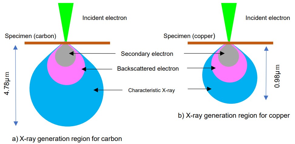
Fig. 1. Generation regions of characteristic X-rays for carbon and copper
The actually measured characteristic X-ray intensities are influenced by various effects: the generated X-rays are absorbed and scattered in the specimen and induce fluorescence excitation. Thus, the depth from which the generated characteristic X-rays can escape the specimen (called “escape depth”), is different depending on these effects. To determine accurate elemental concentrations, it is necessary to apply the quantitative corrections such as ZAF correction and PRZ correction to account for the X-ray attenuation caused by those effects.
Generation process of characteristic X-rays by electron beam irradiation
Fig. 2 illustrates the generation process of characteristic X-rays in a Cu atom irradiated by an electron beam. When an incident electron collides with an inner-shell electron, the impacted electron is ejected from the atom. Then, a vacancy or a hole is created in an inner shell orbital making the atom unstable. To return to the stable state, an electron at a higher energy orbital transitions to the hole created. At this event, a characteristic X-ray is emitted with the energy difference between the two orbitals.
When an electron transitions from the L shell to the K shell, the characteristic X-ray is called the Kα line (Fig. 3a). For the transition from the M shell to the K shell, the X-ray is called the Kβ line (Fig. 3b), and for the transition from the M shell to the L shell, it is called the Lα line (Fig. 3c). The characteristic X-rays are named according to the electron orbitals involved in their emission.
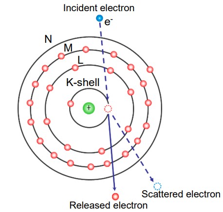
Fig. 2. Generation process of characteristic X-ray for Cu
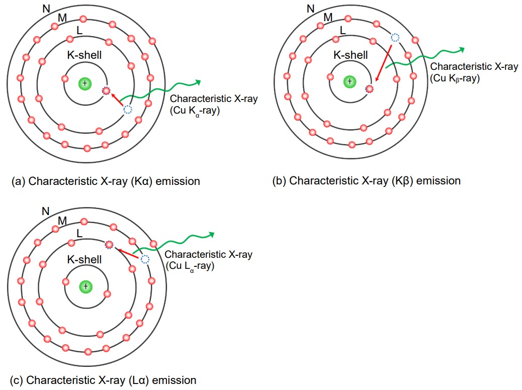
Fig. 3. Characteristic X-ray (Kα, Kβ, Lα) emissions for Cu
Conditions for incident (landing) voltage to obtain sufficient characteristic X-ray intensity
To generate characteristic X-rays, the incident electrons must have an energy equal to or greater than the minimum required to eject an inner-shell electron, known as the absorption-edge. For example, the K absorption edge of Cu is 8.979 keV; thus theoretically, an incident (landing) voltage of 9 kV or higher is sufficient. However, to obtain a spectrum with adequate characteristic X-ray intensity, it is common to set the landing voltage about twice the characteristic X-ray energy of the target element. In the case of Cu, a landing voltage of 15 kV is sufficient to obtain a high intensity spectrum of the Kα line (8.04 keV) as is seen in Fig. 4.
Fig. 5 presents how the energy of the characteristic X-rays (Kα and Lα lines) increases with atomic number. The graph indicates that a landing voltage of 15 kV is sufficient to excite the characteristic X-rays of almost all the elements. For the specimens composed of lighter elements, sufficiently high-intensity X-ray spectra can be acquired by lowering the landing voltage down to 10 kV or even 5 kV.
Therefore, in elemental analysis, it is important to properly set the landing voltage based on the atomic numbers of the specimen.
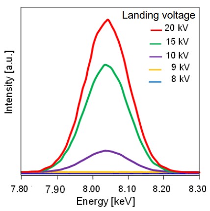
Fig. 4. Dependence of the intensity of Cu-Kα spectrum on landing voltage
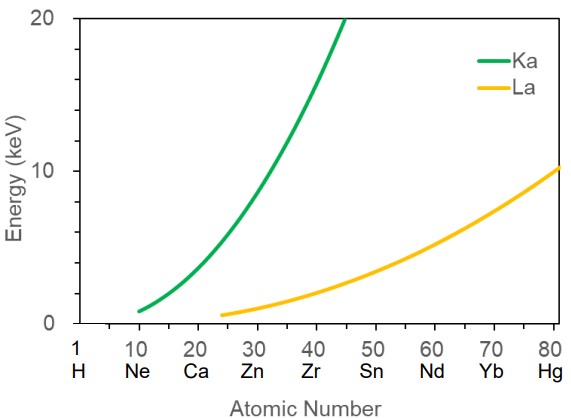
Fig. 5. Dependence of characteristic X-ray energy on atomic number
Related Term(s)
Term(s) with "characteristic X-rays" in the description


Are you a medical professional or personnel engaged in medical care?
No
Please be reminded that these pages are not intended to provide the general public with information about the products.