time-resolved transmission electron microscopy, time-resolved TEM
time-resolved transmission electron microscopy, time-resolved TEM
Time-resolved transmission electron microscopy (time-resolved TEM) is a method to perform time-series imaging for structural change of materials (specimens). In time-resolved TEM, various stimuli (e.g. heating, cooling, electric-magnetic field, tensile stress, laser irradiation) are applied to a specimen, and the resultant phenomena are subjected to time-series observation by using a continuous wave (CW) electron beam or a pulsed electron beam. The correlation between time resolution and spatial resolution in the imaging is shown in a conceptual diagram. In (descending) order of timescales of phenomena, the following three methods are used. (a) A method using a conventional imaging camera (Note 1) at a few seconds (s) to milliseconds (ms), (b) Dynamic TEM (DTEM) at ms to tens of nanoseconds (ns), and (c) Ultrafast TEM (UTEM) at ms to hundreds of femtoseconds (fs).
(a) Method using a conventional imaging camera
When the timescale of structural change of the specimen is slower than the minimum exposure time of a camera (about a few ms), time-resolved imaging can be performed with irradiation of a CW electron beam on the specimen. Time-resolved imaging can be achieved by setting the exposure time of a CMOS camera or a direct electron detection camera and recording the structural changes as a movie. The camera exposure time determines the time resolution. Thus, this method does not require highly-advanced techniques which synchronize stimuli against the specimen with the irradiated electron beam, as described in (b) and (c). For phenomena slower than a few ms, the method can be used for both reversible processes and irreversible processes.
(b) Dynamic TEM (DTEM)
Using DTEM, irreversible processes of structural change of a specimen can be imaged, which occur in the timescale of ms to tens of ns. Since the camera exposure time cannot capture the irreversible structural change in the timescale, a pulsed electron beam is used.
A pulsed electron beam is produced by blanking a CW electron beam using an electrostatic deflector, and by emission of pulsed photoelectrons obtained from irradiation of ultraviolet nanosecond pulsed lasers (wavelength: 250 to 350 nm, etc.) to a Ta cathode. The waiting time between the first and second pulses is called “pulse interval (repetition period)”. The pulse interval and the pulse width are determined by blanking speed (ms to tens of ns) and the type of pulsed laser (microseconds (µs) to tens of ns). To record irreversible changes into the camera with high signal-to-noise ratio, 106 to 108 electrons per pulse are required. The use of ultrahigh power pulsed lasers makes it possible to sustain 106 to 108 electrons even with a very-short pulsed electron beam of tens of ns. The time resolution is determined by the pulse interval and the pulse width.
Since DTEM needs to synchronize generation of a pulsed electron beam, stimuli to the specimen and camera exposure at a precision of ms to tens of ns, more advanced adjustment and data analysis are required than “method using a conventional imaging camera”.
For observing phenomena on the time scale of ms to µs, pulsed electrons blanked by electrostatic deflectors incorporated above the condenser lens are used. For phenomena occurring on timescales below 1 µs, pulsed electrons generated by photoemission from a laser are utilized. The number of pulse electron trains, each containing 10⁶ to 10⁸ electrons per pulse, depends on the laser output, and currently, up to 16 can be generated per 1 µs. In this case, the 16 pulse electron trains that have transmitted through the specimen are deflected in the X-Y direction (two-dimensionally) by electrostatic deflectors inserted above the image observation chamber, and 16 TEM images or electron diffraction patterns (arranged in a 4×4 grid) are recorded sequentially on a single frame of the camera. The size of each of the 16 images is determined by the size of the selected area aperture of the electrostatic deflectors and is set to occupy 1/16 of the single frame. From these 16 images, a frame-by-frame video is generated to observe structural changes in the sample.
(c) Ultrafast TEM (UTEM)
When the timescale of structural change of the specimen reaches ns to fs, the number of electrons per pulse becomes insufficient and thus, it is impossible to observe irreversible change processes. For reversible processes where structural change and relaxation occur repeatedly, the use of UTEM extends the timescale of the observed specimen to ms to fs. That is, UTEM is limited to observe such reversible processes.
In the case of UTEM, a pulsed electron beam is produced by emission of pulsed photoelectrons obtained from irradiation of ultraviolet femtosecond pulsed lasers (wavelength: 250 to 350 nm, etc.) to a Ta cathode, to a LaB6 cathode with a guard ring, or to a W (tungsten) cathode, and by chopping or blanking a CW electron beam using a high-frequency cavity or an electrostatic deflector. UTEM needs to synchronize generation of a pulsed electron beam, stimuli to the specimen and camera exposure at a high precision comparable to the pulse width (e.g. 1 picosecond (ps) to 200 fs).
The time resolution improves as the number of electrons decreases and the pulse width of the electron beam shortens, due to the Boersch effect. In other words, by keeping the laser output low and reducing the number of electrons per pulse to around 1 to 100, it is necessary to minimize the spatial spread (pulse width) of the pulsed electron beam along its propagation direction. In UTEM, the number of incident electrons to the specimen is small and the camera counts decrease. Therefore in some cases of image acquisition, by utilizing high repetition frequencies (kHz to MHz) of pulsed lasers, approximately one millions of images are superposed to improve signal-to-noise ratio.
[Note 1] This method is sometimes called “In-situ TEM”.
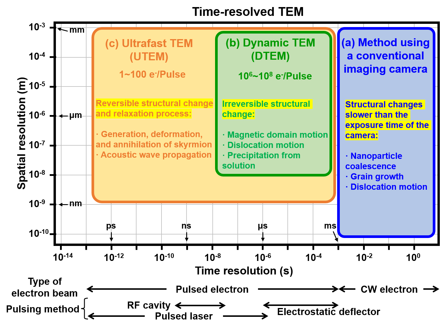
Conceptual diagram: Correlation between time resolution and spatial resolution in imaging
Related Term(s)
Term(s) with "time-resolved transmission electron microscopy, time-resolved TEM" in the description

Are you a medical professional or personnel engaged in medical care?
No
Please be reminded that these pages are not intended to provide the general public with information about the products.


