Serial Section TEM
Serial Section TEM
Serial Section TEM (transmission electron microscopy) is a reconstruction method of the three-dimensional (3D) structure of a specimen using many TEM images of ultra-thin sections prepared serially from a resin-embedded specimen. The method enables us to visualize 3D structures of cells and tissues of a volume of µm3 to mm3 with a resolution of nanometer order.
Fig. 1 shows the procedure of Serial Section TEM. Ultra-thin sections are serially sliced from a resin-embedded specimen using an ultramicrotome, with a 40 to 100 nm thickness of each section. The prepared sections are lined up on a single hole grid with a carbon film fixed, and the images of the same field-of-view of each section are successively taken by TEM. Then, positions of all the TEM images are correctly adjusted, the target structural part (organ) is extracted (called "segmentation"), and by stacking these images the desired 3D structure is reconstructed. Serial Section TEM is a similar method to Array Tomography in SEM (scanning electron microscopy).
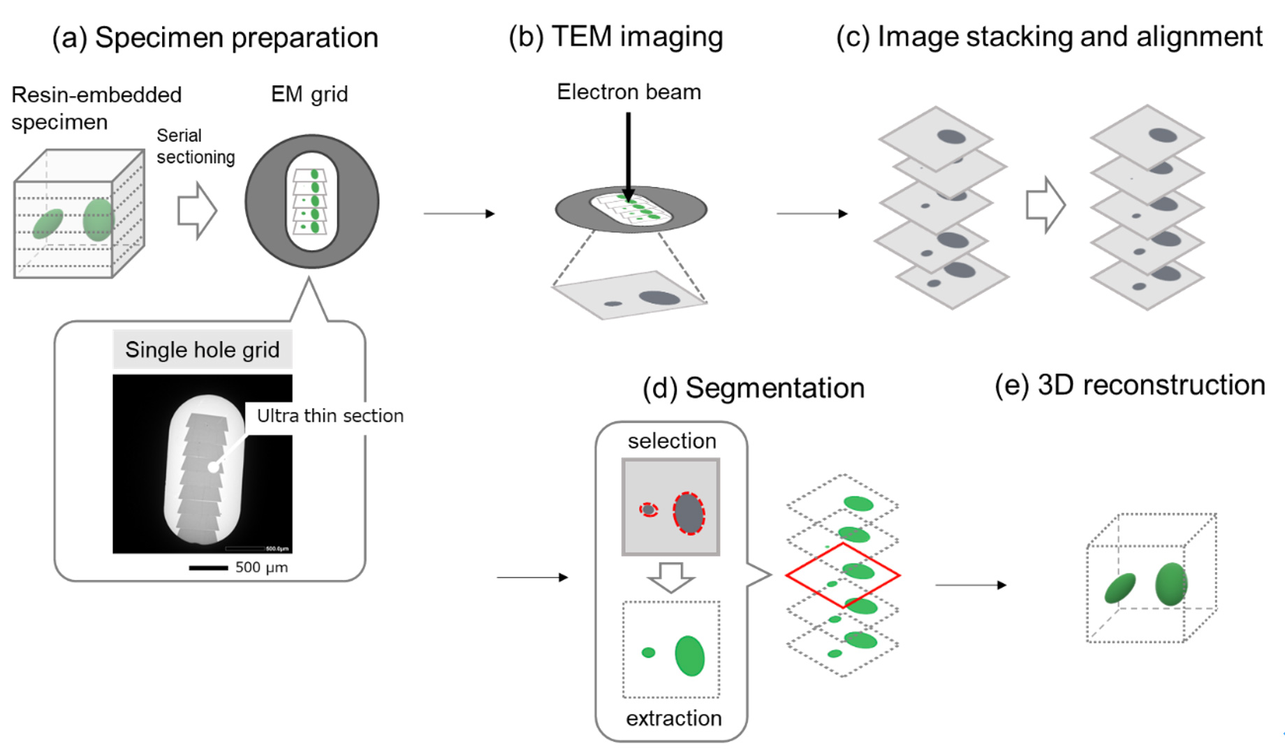
Fig. 1. Procedure of Serial Section TEM
(a) Ultra-thin sections are serially sliced from a resin-embedded specimen using an ultramicrotome, and then the sections are lined up on a single hole grid with a carbon film fixed. Bottom: the aligned ultra-thin sections.
(b) TEM images of the same field-of-view of each section are successively taken.
(c) Positions of all the TEM images are correctly adjusted.
(d) Only the target structural part (organ) is extracted (called "segmentation").
(e) 3D structure of the desired part is reconstructed by stacking these images.
Fig. 2. 3D-reconstructed image of chloroplasts (green) of carrot leaves and cell walls (white).
Many chloroplasts present inside the cell walls are clearly seen.
Size of 3D reconstruction: (x, y, z) = (9.4, 9.4, 12.4) µm, Specimen: carrot leaves, Instrument: JEM-1400Flash, Accelerating voltage: 120 kV, Magnification: x4,000, Number of captured images: 177. For a specimen supporting film, not only a carbon film, but also the other films can be used. In this case, a silicon nitride film (SiN Window Chip) was used.
Related Term(s)
Term(s) with "Serial Section TEM" in the description

Are you a medical professional or personnel engaged in medical care?
No
Please be reminded that these pages are not intended to provide the general public with information about the products.


