Autoradiography (Autoradiography in Electron Microscopy)
Autoradiography (Autoradiography in Electron Microscopy)
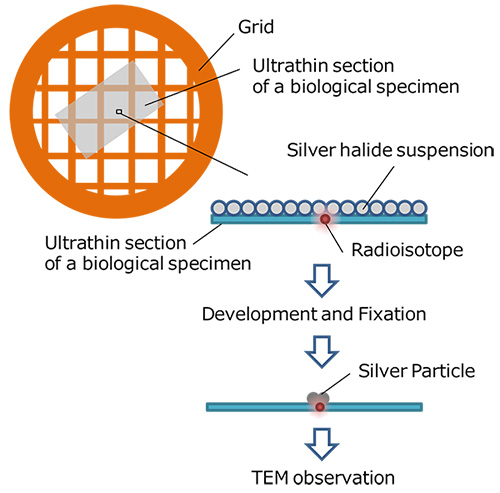
Autoradiography in Electron Microscopy is a method to observe a specific site of a biological specimen labeled with a substance containing a radioactive isotope. The method is implemented in the following procedure.
A substance containing a radioactive isotope is doped into a biological specimen for labeling specific tissues or cells. The biological specimen is thinned down to an ultrathin section and a photo-sensitive emulsion (silver halide suspension) is applied to the thin section. Silver halides in the vicinity of the labeled sites are exposed with β rays emitted from the radioactive isotope. When photo-developed, silver particles are segregated at the labeled sites. When the section is observed with a transmission electron microscope, the positions of the labeled tissues or cells can be identified from the localized silver particles.
In order to perform high-resolution observation of the sites of the labeled tissues or cells, tritium (which emits small energy β-rays) is often used as a radioactive isotope because tritium causes small silver segregates in the photosensitive emulsion.
An example of Autoradiography in Electron Microscopy: Thymidine containing radioactive tritium is applied to label the sites where cell divisions are active in a biological specimen. The labeled sites are revealed from the segregated silver particles by observing the electron microscope image of the specimen.
Related Term(s)
Term(s) with "Autoradiography (Autoradiography in Electron Microscopy)" in the description

Are you a medical professional or personnel engaged in medical care?
No
Please be reminded that these pages are not intended to provide the general public with information about the products.


