axial geometrical aberration
axial geometrical aberration
The geometrical aberrations (departures of the path of electron beams from the path of ideal imaging on the image plane) are expressed as a function of r (distance of the electron beam from the optical axis) and α (opening angle between the electron beam and the optical axis). Among those aberrations, aberrations which depend only on the opening angle α, are called the axial geometrical aberrations (hereinafter, referred to be “axial aberrations”). It should be noted that, aberrations depending on r, and on both r and α, are called the off-axial geometrical aberrations (hereinafter, referred to be “off-axial aberrations”).
Table 1 lists the axial aberrations. The axial aberrations are classified by the order n of the opening angle α and the symmetry in the azimuth direction. For example, the first-order axially-symmetrical aberration is called “defocus” and the first-order two-fold symmetrical aberration is called “two-fold astigmatism.” The third-order axially-symmetrical aberration is called “spherical aberration”, which is well known as one of Five Seidel aberrations. Aberrations that are left blank in Table 1, such as the second-order two-fold symmetrical aberration, do not appear in principle.
When the axial aberrations exist, electron beams do not come to a single point on the image plane, but broaden to reduce the resolution of the image (Fig. 1). Among the many lenses of a transmission electron microscope (TEM), the aberration of the objective lens (post-field) has the greatest effect on the resolution of the image because the opening angle α is the greatest for the electron beam passing through the objective lens located closest to the specimen (Fig. 2). When observing the image of a specimen at a high magnification (high resolution electron microscopy), the observation area is small or the distance r is small. Thus, the off-axial aberrations can be ignored and only the axial aberrations of the objective lens are taken in account.
On the other hand, for obtaining a high-resolution image using scanning transmission electron microscopy (STEM), it is needed to converge the incident electron beam (incident probe) onto a specimen as smallest as possible. Thus, it is important to reduce the axial aberration of the condenser lens (actually the pre-field of the objective lens) located just in front of the specimen.
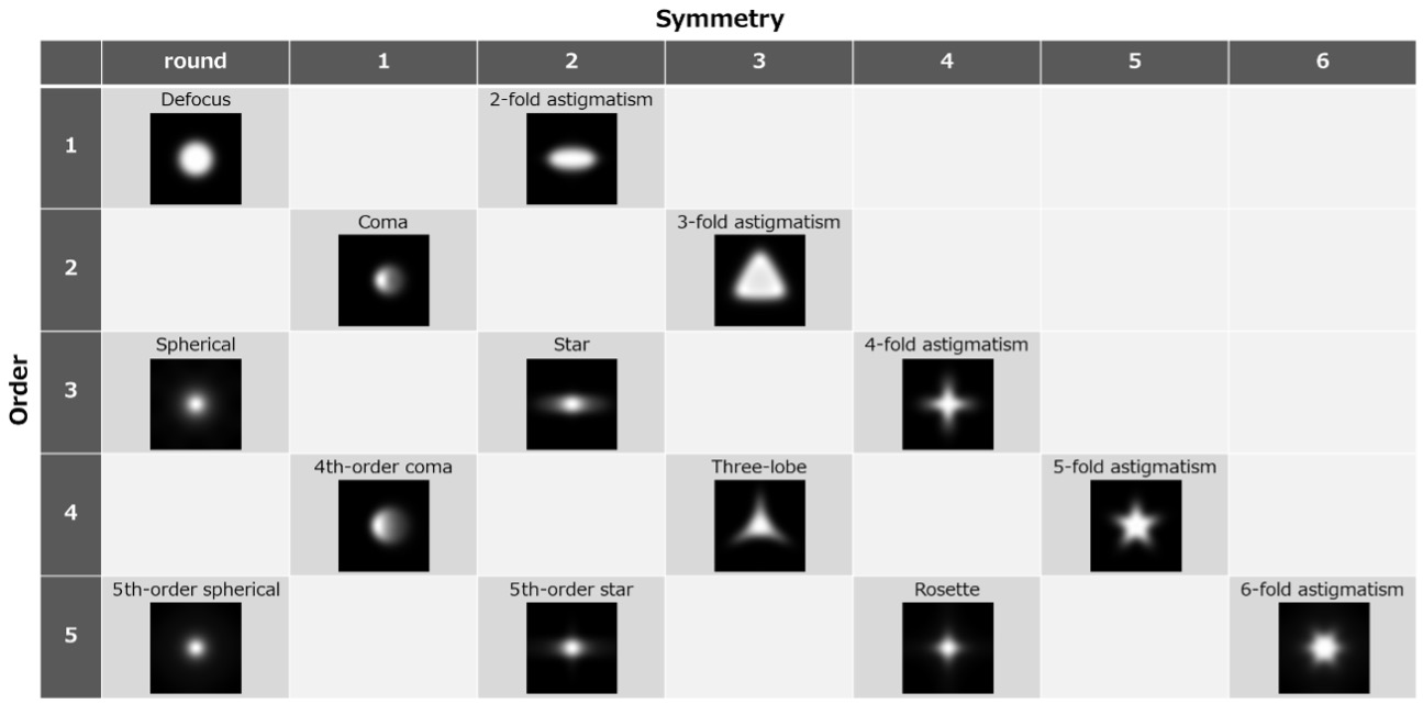
Table 1. List of axial geometrical aberrations. The aberrations are classified by giving the order n of the opening angle α in the vertical direction and the symmetry in the azimuthal direction in the horizontal direction. The blurring of the electron probe in STEM due to each aberration is shown.
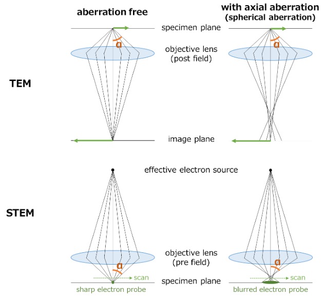
Fig. 1. Ray paths through the objective lens with (right) and without (left) the axial geometrical aberrations in TEM (top) and STEM (bottom). In TEM, the aberration causes the electrons coming from each point of a specimen blurring on the image plane. In STEM, the aberration causes the electrons emitted from a (virtual) electron source a beam-broadening on the specimen plane. When an image is formed by scanning the specimen using the broadened beam (probe), the image is blurred.
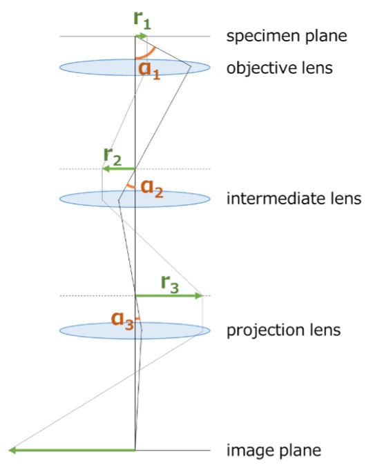
Fig. 2. The increase of the distance r of the electron beam from the optical axis and the decrease of the opening angle α between the electron beam and the optical axis when an image is magnified by several lenses in the imaging system of the microscope. The more the image is magnified, the greater the distance r (r1 < r2 < r3), and to the contrary, the smaller the angle α (α1 > α2 > α3). Since the axial aberrations depend only on the opening angle α, the aberration in the upper lens (objective lens) has a crucial effect on degrading the image.
Appendix: The axial geometrical aberrations and advancement of correction of the aberrations
The spherical aberration of the objective lens (third-order axially-symmetrical aberration) had long been limiting the resolution of the image of the transmission electron microscope.
In the 1980s, a unique method was developed to cancel out the image blur caused by spherical aberration by adjusting the amount of defocus (Scherzer defocus) of the objective lens, which enabled us to take high-resolution images (~0.2 nm) using an electron beam with an angular range of approximately 10 mrad.
Since the late 1990s, spherical aberration correctors of the hexapole and quadrupole-octupole types have been put into practical use, enabling spherical aberration coefficients (Cs) to be reduced to zero. The spherical aberration corrector, Cs corrector, can correct not only the spherical aberration, but also the axial coma aberration and parasitic aberrations including the 3-fold astigmatism, star aberration, and 4-fold astigmatism. With this breakthrough, an electron beam can be converged onto a single point on the image plane even when an electron beam with a large opening angle α (semi-angle ~30 mrad) is accepted. In the early 2000s, practical use of the Cs corrector achieved the image resolution better than 0.1 nm. Furthermore in the late 2000s, the image resolution reached better than 0.05 nm owing to the improvement in the stability of the microscope and development of a cold field emission gun (which improved monochromaticity of the electron source).
In sextupole Cs correctors of the early 2000s, the angular range of aberration correction was limited to ~30 mrad by the six-fold astigmatism. But, in the late 2000s, aberration correctors (opening angle ~60 mrad) which can correct the six-fold astigmatism, put into practical use. By using an electron beam with an opening angle of ~40 mrad or better, an atomic resolution of about 0.1 nm was achieved at low acceleration voltages of 30 to 60 kV.
This facilitates to observe monoatomic layers such as graphene. It is important to note that the increase of the opening angle contributes to improve the resolution of the image in the depth direction because the focal depth is inversely proportional to the square of the opening angle.
Related Term(s)
Term(s) with "axial geometrical aberration" in the description

Are you a medical professional or personnel engaged in medical care?
No
Please be reminded that these pages are not intended to provide the general public with information about the products.


