Ronchigram
Ronchigram
A Ronchigram is a projection image (pattern) of a specimen formed on the diffraction plane with a convergent incident electron beam focused near the specimen using a probe-forming lens. The Ronchigram enables us to determine the optical characteristics (amount of aberration) of the electron probe formed near the specimen using the probe-forming lens. The image is used to obtain the exact focus of the incident electron probe onto the specimen in STEM observations, to know the angular range of the electron probe with no aberration, and to compensate the axial astigmatism.
When the convergence point of the incident electron probe becomes closer to an amorphous specimen, the magnification of the specimen-shadow image (pattern) seen in the Ronchigram is increased. When the convergence point of the probe is exactly focused onto the specimen, the magnification of the Ronchigram or the shadow image becomes infinite and its intensity becomes uniform. By adjusting the Ronchigram to have a uniform intensity, the exact focus of the incident beam onto the specimen is confirmed. Measurement of the angular range of the area with a uniform intensity in the Ronchigram allows us to determine the angular range of the incident probe with no aberration or to know the quality of the electron-probe. If the third-order spherical aberration is not corrected, the area with a uniform electron intensity is confined to a small extent. When the aberration is corrected, the area is largely extended. This means that the angular range of the electron beam focused onto a point of the specimen is expanded due to vanishing of the aberration.
The Ronchigram obtained using a crystalline specimen exhibits interference fringes, when the incident beam with an incidence angle larger than the diffraction angle from a lattice plane of the crystal is illuminated onto the specimen with a small defocus. Appearance of the interference fringes means that the probe diameter on the focal plane is smaller than the lattice spacing. From this experiment, the probe size on the focal plane used for STEM observations can be known.
The term “Ronchigram” is named after V. Ronchi, who proposed the method to originally examine the performance of a lens of light optics.
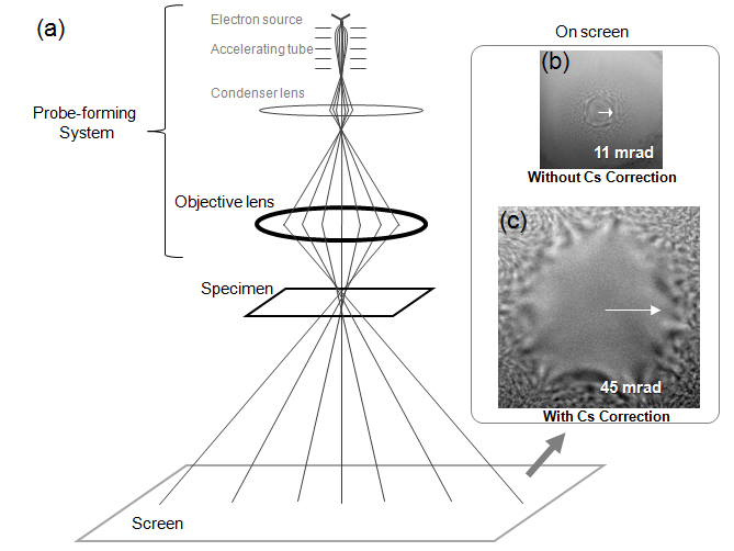
Fig. (a) Ray diagram of Ronchigram.
Fig. (b), Fig. (c) Ronchigram patterns obtained from an amorphous thin-film specimen. In the case of no aberration correction (Fig. (b)), the area with a uniform electron intensity is confined to a small extent (semi-angle: ~11 mrad). When the aberration is corrected (Fig. (c)), the area is largely extended (semi-angle: ~45 mrad).
When the convergence point of the incident electron probe becomes closer to an amorphous specimen, the magnification of the specimen-shadow image (pattern) seen in the Ronchigram is increased. When the convergence point of the probe is exactly focused onto the specimen, the magnification of the Ronchigram or the shadow image becomes infinite and its intensity becomes uniform. By adjusting the Ronchigram to have a uniform intensity, the exact focus of the incident beam onto the specimen is confirmed. Measurement of the angular range of the area with a uniform intensity in the Ronchigram allows us to determine the angular range of the incident probe with no aberration or to know the quality of the electron-probe. If the third-order spherical aberration is not corrected, the area with a uniform electron intensity is confined to a small extent. When the aberration is corrected, the area is largely extended. This means that the angular range of the electron beam focused onto a point of the specimen is expanded due to vanishing of the aberration.
The Ronchigram obtained using a crystalline specimen exhibits interference fringes, when the incident beam with an incidence angle larger than the diffraction angle from a lattice plane of the crystal is illuminated onto the specimen with a small defocus. Appearance of the interference fringes means that the probe diameter on the focal plane is smaller than the lattice spacing. From this experiment, the probe size on the focal plane used for STEM observations can be known.
The term “Ronchigram” is named after V. Ronchi, who proposed the method to originally examine the performance of a lens of light optics.
Fig. (a) Ray diagram of Ronchigram.
Fig. (b), Fig. (c) Ronchigram patterns obtained from an amorphous thin-film specimen. In the case of no aberration correction (Fig. (b)), the area with a uniform electron intensity is confined to a small extent (semi-angle: ~11 mrad). When the aberration is corrected (Fig. (c)), the area is largely extended (semi-angle: ~45 mrad).
Related Term(s)
Term(s) with "Ronchigram" in the description

Are you a medical professional or personnel engaged in medical care?
No
Please be reminded that these pages are not intended to provide the general public with information about the products.


