bright-field image
bright-field image
An image that is produced by the transmitted wave (the wave that undergoes no diffraction) in a diffraction pattern formed on the back focal plane of the objective lens, using the objective aperture. In the image, a location where diffraction takes place appears dark, whereas a location where diffraction does not take place appears bright. The bright-field image, together with the dark-field image, is used for analysis of lattice defect and measurement of specimen thickness.
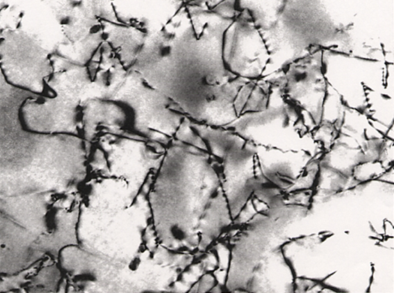
Fig. Bright-field image of lattice defects (dislocation lines) in an FeAl alloy. The image was taken in such a way that the distorted area caused by the dislocations does not satisfy the Bragg diffraction condition. Thus, the dislocation lines appear dark. The zigzag contrast of the dislocation lines is created by a dynamical diffraction effect, which depends on the depth of the dislocations in the specimen.
Fig. Bright-field image of lattice defects (dislocation lines) in an FeAl alloy. The image was taken in such a way that the distorted area caused by the dislocations does not satisfy the Bragg diffraction condition. Thus, the dislocation lines appear dark. The zigzag contrast of the dislocation lines is created by a dynamical diffraction effect, which depends on the depth of the dislocations in the specimen.
Related Term(s)
Term(s) with "bright-field image" in the description

Are you a medical professional or personnel engaged in medical care?
No
Please be reminded that these pages are not intended to provide the general public with information about the products.


