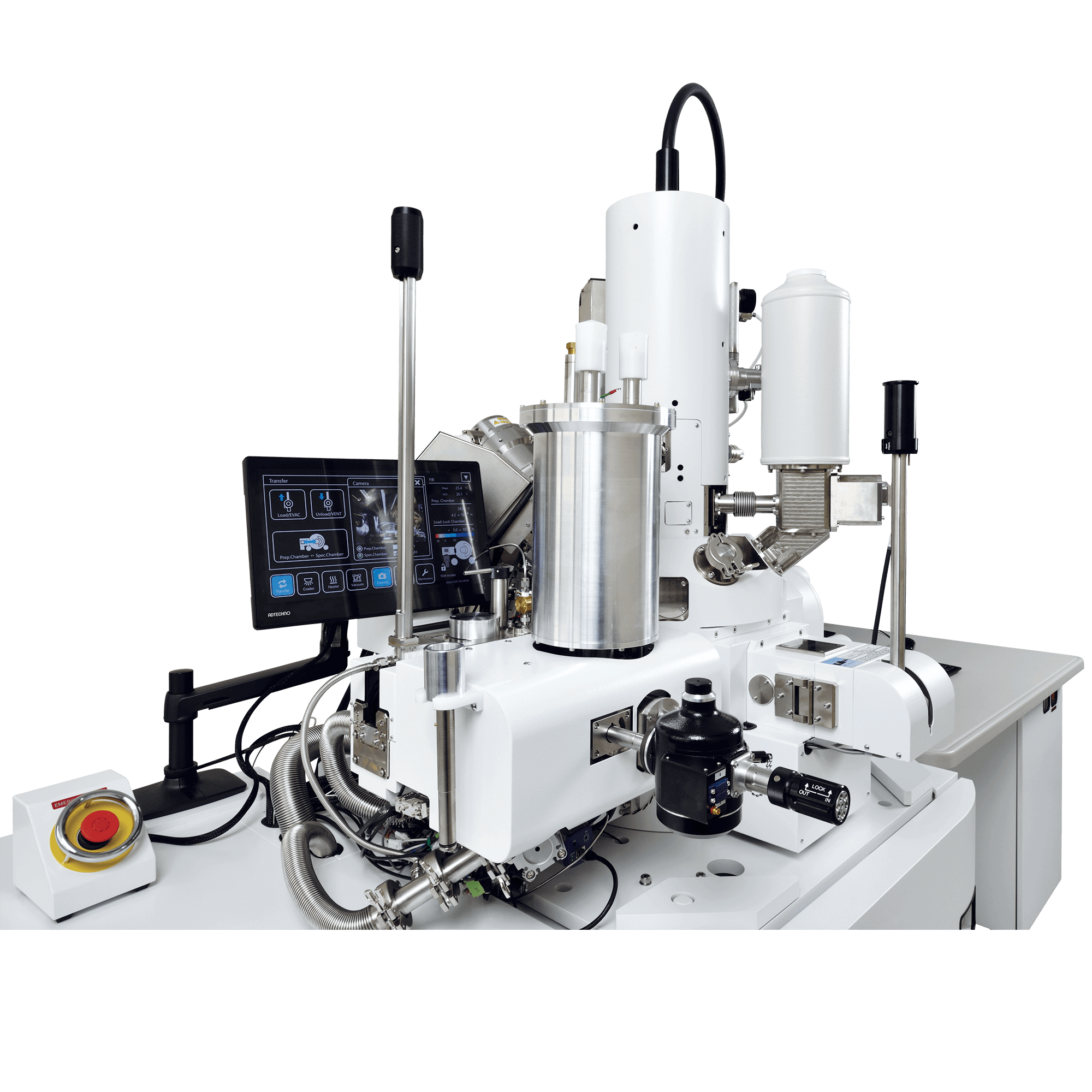
CRYO ARM™ 300 (JEM-Z300FSC)
Field Emission Cryo-Electron
Microscope
 Powered by Bioz
The JEM-Z300FSC (CRYO ARM™ 300), equipped with a cold field emission gun, an in-column Omega energy filter, a side-entry liquid nitrogen cooling stage and an automated specimen exchange system, is a cryo-electron microscope (cryo-EM) that enables observation of bio-molecules at cryo-temperature. The automated specimen exchange system features the storing of up to 12 samples. In addition, the system allows for the exchange of an arbitrary one or more samples, thus enabling flexible scheduling. Furthermore, the combined use of a newly-designed in-column Omega energy filter and a Hole-free phase plate dramatically enhances the contrast of TEM images of biological specimens.
Features
Automated specimen exchange system
The system is composed of a specimen stage to cool samples to liquid nitrogen temperature and a cryo-transfer system to automatically transfer the cooled samples to the cryo-stage. Liquid nitrogen is automatically supplied to the liquid nitrogen tank as required. This automated system features the storing of up to 12 samples and the exchange of an arbitrary one or more samples while the rest of the samples are kept cooled between the specimen stage and specimen exchange system.
Cold field emission gun (Cold FEG)
A Cold FEG produces a high-brightness electron beam with very small energy spread, offering high coherency. Thus, the CRYO ARM™ 300 achieves high resolution, high contrast imaging.
In-column Omega energy filter
Equipped with an in-column Omega energy filter, the CRYO ARM™ 300 acquires energy filtered images and energy loss spectra. Zero-loss images acquired with the microscope provide high contrast with reduced chromatic aberration.
Automated image acquisition software for Single Particle Analysis
The CRYO ARM™ 300 incorporates automated software. The software allows for automated detection of holes on the specimen grid for efficient acquisition of Single Particle Analysis images.
Hole-free phase plate *1
This unique phase plate is suitable for higher contrast in biological specimens that originally provide only low contrast.
Auto adjustment functions *2
Auto focus, auto coma-free alignment, auto parallel-beam illumination and other automated adjustments are available, enabling image acquisition under optimum conditions.
Optional unit.
Images acquired with the bottom mount camera are used.
Research Publications Using JEM-Z300FSC
Specifications
Main instrument
| Electron gun | Cold field emission gun (Cold FEG) |
|---|---|
| Accelerating voltage | 300kV |
| Energy filter | In-column Omega energy filter |
| Maximum specimen tilt angle | ± 70° |
Specimen Stage / Automated specimen exchange system
| Specimen stage | |
|---|---|
| Coolant | Liquid nitrogen Automated liquid-nitrogen filling system built-in |
| Specimen cooling temperature | 100K or less |
| Temperature measurement position | Specimen, Cryo-shield, LN2 tank |
| Specimen movements | |
| X、Y | Motor drive (movements: ±1 mm) Piezoelectric elements (movements: ±0.5 μm) |
| Z | Motor drive (movements: ±0.2 mm) |
| Tilt-X | Motor drive (tilts: ±70°) |
| Rotation within the specimen plane | 0° or 90° |
| Specimen exchange system | Air-lock Automated cryo-transfer system built-in |
| Cooling temperature (specimen exchange chamber) |
105K or less |
| Specimen exchange cartridge | Up to 4 specimens can be changed at one time. |
| Specimen parking stage | Up to 12 specimens can be held. |
Catalogue Download
CRYO ARM™ 300 (JEM-Z300FSC) Field Emission Cryo-Electron Microscope
Application
Application JEM-Z300FSC
Technical Development of Electron Cryomicroscopy and Contributions to Life Sciences
Gallery
Reference
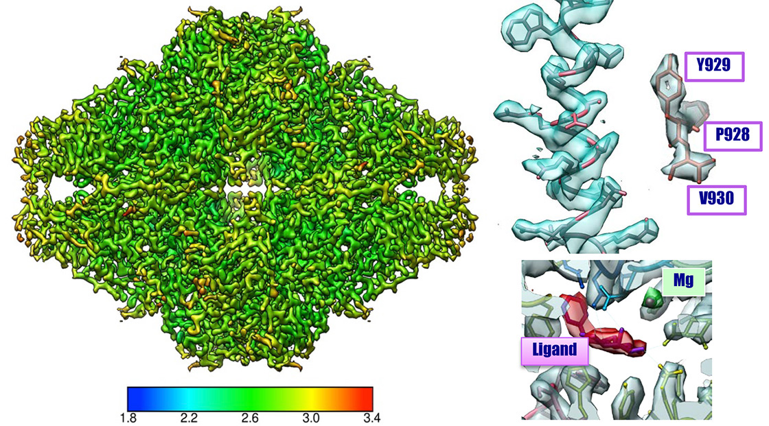
Innexin-6 gap junction channel
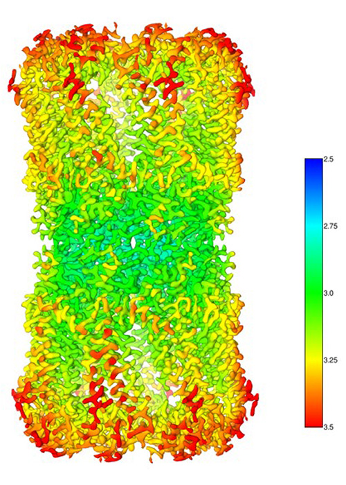
Experimental conditions
Sample: Innexin-6 (Caenorhabditis elegans)
Microscope: CRYO ARM™300 (300kV CFEG) with Gatan K2
Software used for image acquisition : JADAS (1974 images)
Number of particles: 91,613 (Initial pickup), 37,767 (for 3D reconstruction)
Software used for image analysis: Relion3
Resolution: 3.0 Å (at FSC = 0.143)
Sample by courtesy of Prof. A. Oshima (Nagoya University)
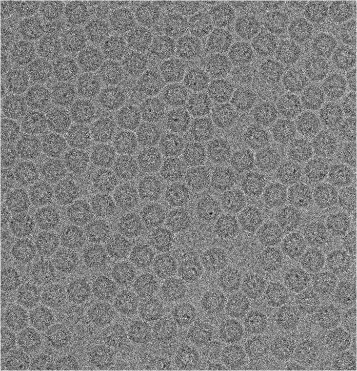
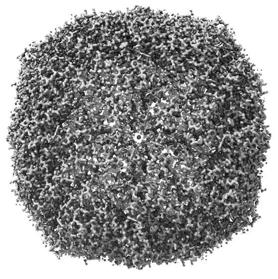
Apoferritin
The highest resolution 1.53 Å achieved by cryoEM 2019.02
mouse apoferritin
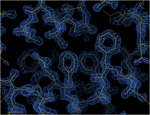
Phenylalanine
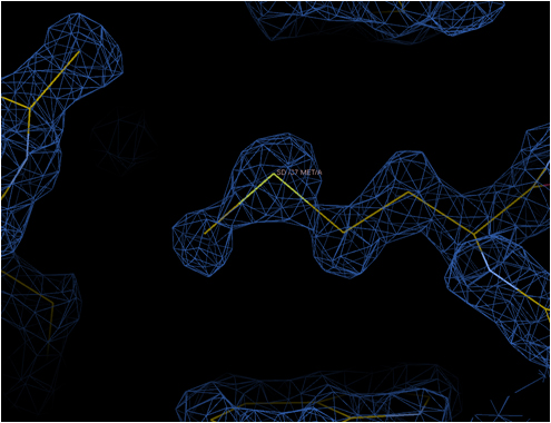
Methionine sulfur atom
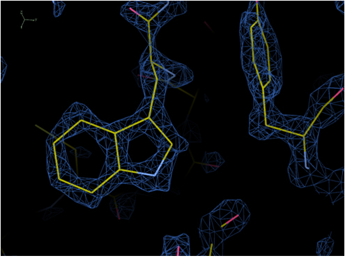
Tryptophan
Optics features: Cold FEG 300 kV & Ω-type energy filter with 20 eV slit width
Detector: Gatan K2 (image pixel size: 0.495 Å, mag x100,000)
Grid: Quantifoil 1.2/1.3 Cu 200 mesh, kept in the autoloader for 3 days before data collection
No. of micrographs: 974 collected over 24 h, 840 used for image analysis
No. of particles: 120,295 used for final reconstructionSoftware: RELION 3.1b, CTFFIND4
Software: RELION 3.1b, CTFFIND4
Resolution: 1.53 Å (B-factor: 47)
Note: the first 56 images alone produced a map of 1.76 Å resolution (B-factor: 45)
Mouse apoferritin plasmid from Yanagisawa, Danev & Kikkawa @Tokyo University
Kato, Makino, Nakane, Terahara, Kaneko, Shimizu, Motoki, Ishikawa, Yonekura & Namba 2019.02 (EMDB-9865)
ß-galactosidase
β-galactosidase 2.43 Å resolution CRYO ARM™
Three-dimensional calibration image of β-galactosidase obtained using a cryo-electron microscope (CRYO ARM™ 200) with an accelerating voltage of 200 kV
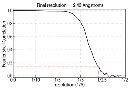
Sample:
β-galactosidase with PETGMicroscope:
CRYO ARM™ (Schottky 200 kV) / K2 summitNumber of Images:
2,500 over 3 days by JADASImage pixel size:
0.8 Å/pixelNumber of particle images:
350,000(Initial pickup), 88,564 (for final 3D reconstruction)Software:
Motioncor2, Gctf, Gautomatch, Relion2.0Total dose:
70 e-/Å2 (70 frames (0.2 sec/frames x 14 sec)
Data: courtesy by Dr. T. Kato and Dr. K. Namba, Osaka University, August 2017
Related Products
CRYO-FIB-SEM CryoLameller
This CRYO-FIB-SEM system incorporates a liquid nitrogen cooling stage and a cryocooled specimen transfer mechanism for frozen specimens, making it possible to prepare TEM specimens such as biopolymers.The specimen transfer mechanism has a built-in sputter coating function. Therefore, this CRYO-FIB-SEM system alone can perform a series of processes to create TEM specimens from frozen specimens including conductivity coating, protective film forming, and FIB processing.In addition, by using JEOL's CRYO ARM™ cartridge, direct specimen transfer to the CRYO ARM™ after TEM specimen preparation becomes easier.
JEM-120i Electron Microscope
Transmission Electron Microscopes(TEM) with 120kV accelerating voltage are widely used in soft material fields such as biology and polymer. We newly developed JEM-120i with the concept of "Compact", "Easy To Use", and "Expandable". With the new external appearance, this instrument has evolved into a useful tool that anyone can use easily, from operation to maintenance.
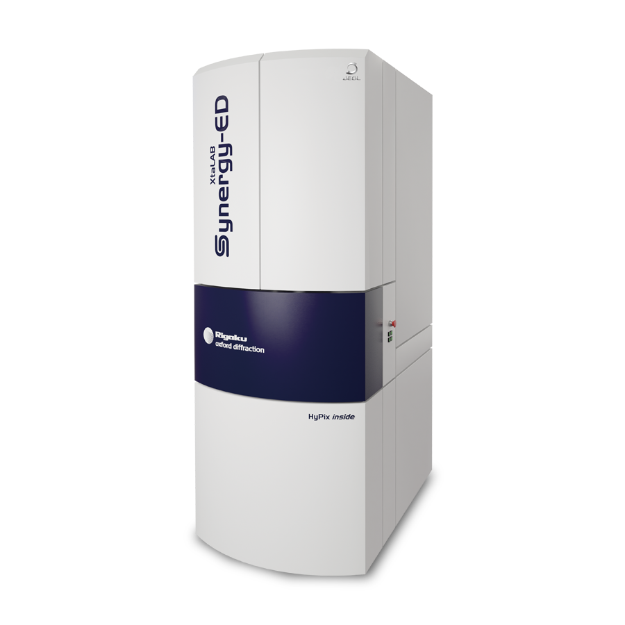
Electron Diffractometer XtaLAB Synergy-ED
XtaLAB Synergy-ED is a completely new electron diffractometer born from the synergy of Rigaku and JEOL's core technologies.It integrates Rigaku's high-speed, ultra-sensitive detector HyPix-ED, the "CrysAlisPro for ED" software that covers everything from measurement to structural analysis, and JEOL's electron beam generation and control technologies that have been refined over many years. By integrating the flow from selection of measurement samples (nanocrystals) to data collection and analysis, electron diffraction structure analysis can be easily used by non-specialists who lack the expertise in electron microscopy and crystallography that is conventionally required.
More Info
Are you a medical professional or personnel engaged in medical care?
No
Please be reminded that these pages are not intended to provide the general public with information about the products.