JEM-2200FS
Field Emission Electron
Microscope
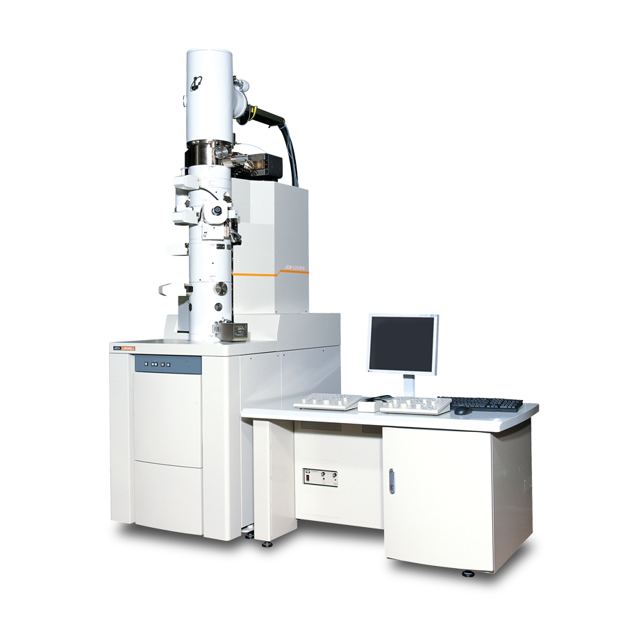
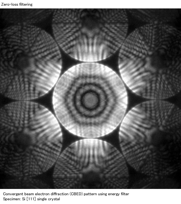
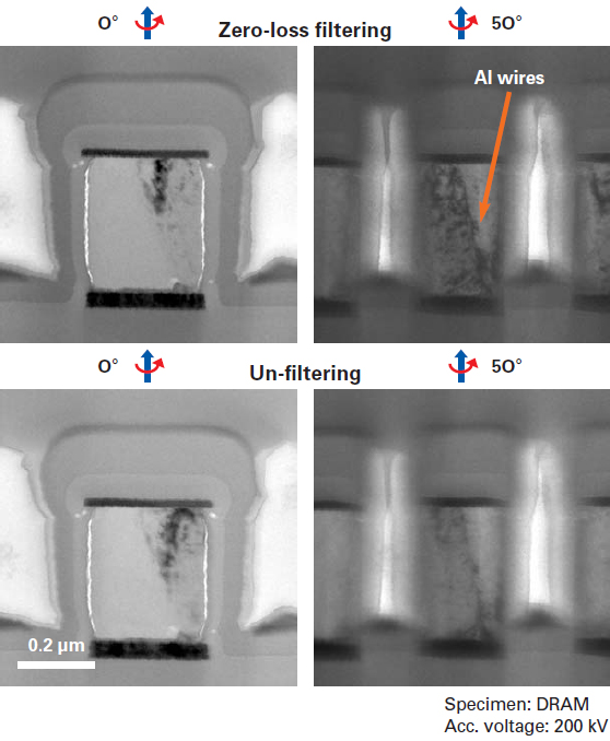
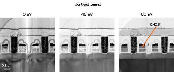
The JEM-2200FS, a state-of-the art analytical electron microscope, is equipped with a 200kV field emission gun (FEG) and the in-column energy filter (Omega filter) that allows a zero-loss image, where inelastic electrons is eliminated, resulting in clear images with high contrast. And energy-filtered images forming with electrons at low loss or core loss energy provide chemical state or elemental information of a sample. Also, spectroscopy for elemental analysis and chemical analysis of specimens is available.
Features
In-column energy filter (Omega filter)
The in-column energy filter enables you to obtain energy-filtered images and electron energy loss spectra. The optimally designed filter provides distortion-free filtered images.
Control system
The main components of the JEM-2200FS, such as the optical system, goniometer stage and evacuation system, are fully PC-controlled. This system stably produces high-quality data.
Imaging system
A new imaging system, consisting of four-stage intermediate lenses and two-stage projector lenses, achieves rotation-free energy-filtered TEM images and diffraction patterns over a wide range of magnifications and camera length.
Piezo-controlled goniometer
A new goniometer stage that incorporates a piezo device offers smooth operation for searching fields of view at the atomic level.
Integration to other instruments
The microscope can be fully controlled with PC. The design concept enables us to integrate EDS system and CCD cameras.
Specifications
| Configuration※1 | Ultrahigh resolution (UHR) |
High resolution (HR) |
High specimen tilt (HT) |
Cryo (CR) |
High Contrast (HC) |
|---|---|---|---|---|---|
| Resolution | |||||
| Point Lattice |
0.19 nm 0.1 nm |
0.23 nm 0.1 nm |
0.25 nm 0.1 nm |
0.27 nm 0.14 nm |
0.31 nm 0.14 nm |
| Energy Resolution | 0.8 eV(zero-loss FWHM) | ||||
| Acc. Voltage | 160 kV,200 kV※2 | ||||
| Minimum step size Energy shift |
50 V 3,000 V maximum (in 0.2 V steps) |
||||
| Electron source | |||||
| Emitter | ZrO/W(100) Schottky | ||||
| Brightness | ≧4×10⁸ A/cm2 ・ sr | ||||
| Vacuum | ×10-8 Pa order | ||||
| Probe current | 0.5 nA at 1 nm probe | ||||
| Power Stability | |||||
| Acc. voltage | ≦1×10⁻⁶/min | ||||
| OL current | ≦1×10⁻⁶/min | ||||
| Filter current | ≦1×10⁻⁶/min | ||||
| Objective Lens | |||||
| Focal length | 1.9 mm | 2.3 mm | 2.7 mm | 2.8 mm | 3.9 mm |
| Spherical aberration |
0.5 mm | 1.0 mm | 1.4 mm | 2.0 mm | 3.3 mm |
| Chromatic aberration |
1.1 mm | 1.4 mm | 1.8 mm | 2.1 mm | 3.0 mm |
| Minimum step | 1.0 nm | 1.4 nm | 1.8 nm | 2.0 nm | 5.2 nm |
| Spot Size (diameter) | |||||
| TEM mode | 2 to 5 nm | 7 to30 nm | |||
| EDS mode | 0.5 to 2.4 nm | 1.0 to2.4 nm | - | 4 to20 nm | |
| NBD mode | - | - | |||
| CBD mode | 1.0 to2.4 nm | - | |||
| CB Diffraction | |||||
| Convergence angle(2α) |
1.5 to 20 mrad or more | - | |||
| Acceptance angle | ±10 ° | - | |||
| Magnification | |||||
| MAG mode | ×2,000 to 1,500,000 | ×2,000 to 1,200,000 | ×1,500 to1,000,000 | ×1,200 to600,000 | |
| LOW MAG mode | ×50 to1,500 | ||||
| SA MAG mode | ×10,000 to800,000 | ×8,000 to 600,000 | ×8,000 to500,000 | ×5,000 to 400,000 | |
| Field-of view size for energy selected image | 80 mm dia. on final image plane (film) when 10 eV is selected 25 mm dia. on final image plane (film) when 2 eV is selected |
||||
| Camera Length | |||||
| SA diffraction | 150 to1,500mm | 200 to2,000 mm | 250 to2,500 mm | 300 to3,000 mm | |
| EELS Dispersion | |||||
| On slit | 1.2 μm/eV at 200 kV | ||||
| On final image plane | 50 to300 μm/eV at 200 kV | ||||
| Specimen chamber | |||||
| Shift(XY / Z) | 2mm/0.2mm | 2mm/0.4mm | 2mm/0.4mm | 2mm/0.4mm | 2mm/0.4mm |
| Specimen tilt X / Y *3 | ±25°/±25° | ±35°/±30° | ±42°/±30° | ±15°/±10° | ±38°/±30° |
| Specimen tilt angle(X)※4 | ±25° | ±80° | ±80° | ±80° | ±80° |
| EDS※5 | |||||
| Solid angle※6 | 0.13 sr | - | 0.09 sr | ||
| Take-off angle | 25° | - | 20° | ||
Select either configuration when ordering the JEM-2200FS.
80 kV, 100 kV and 120 kV become possible when using the optional short-circuit switches for the accelerating tube.
When the Specimen Tilting Holder is used.
When the High Tilt Specimen Retainer is used.
An optional EDS detector is needed.
When 30 mm2 detector is installed.
Catalogue Download
JEM-2200FS Field Emission Electron Microscope
Application
Application JEM-2200FS
Visualization of hydrogen-bonding: Electron and NMR nano-crystallography
Exploring Biological Samples in 3D Beyond Classic Electron Tomography
Atomic-Resolution Elemental Mapping by EELS and XEDS in Aberration Corrected STEM
Gallery
Related Products
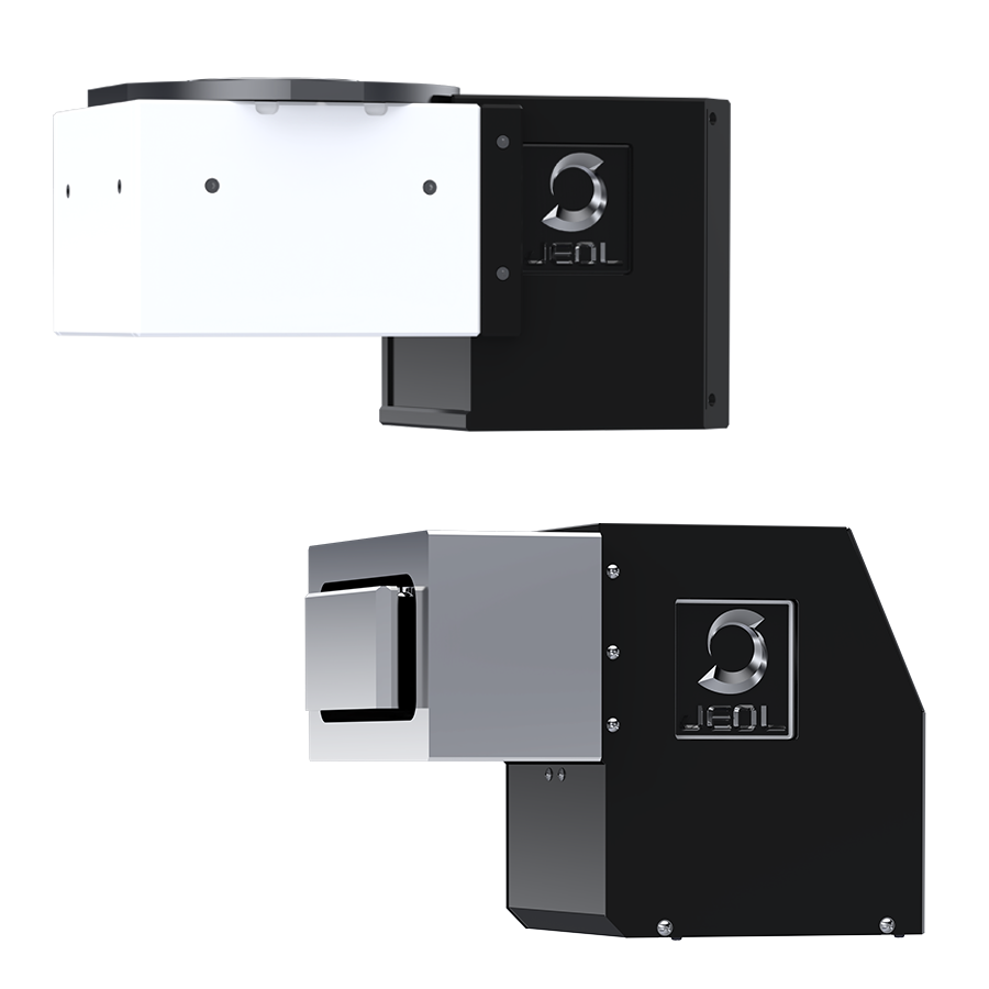
SightSKY™ Series High-Sensitivity, Low-Noise Fiber-Coupling CMOS Camera
High-sensitivity, low-noise 19 M pixel CMOS sensor enables imaging of fine specimen detail and obtains high signal to noise images even at low electron doses.A Global shutter and high frame rate (58 fps/full pixel mode) enable image series acquisitions with less artifacts during in-situ dynamic observation studies.The SightSKY™ camera system offers integration with JEOL’s FEMTUS™ Integrated Analysis Platform, providing user-friendly operation and data acquisition.
More Info
Are you a medical professional or personnel engaged in medical care?
No
Please be reminded that these pages are not intended to provide the general public with information about the products.

