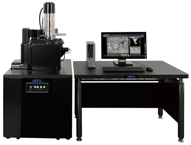
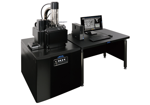
【DISCONTINUED】JSM-IT300 InTouchScope™ Scanning Electron Microscope
DISCONTINUED
This product is no longer available.
If you would like to know the latest information about your preferred product or to find out more about alternatives, please click on the link below. We hope you will continue to use our products.
In addition to the high image quality observation due to the improvement of the illuminating system, the vacuum system and the signal processing system, the JSM-IT300 is a Scanning Electron Microscope, which can be operated with a high throughput by using touch panel operation and the high speed stage.
Features
Advanced Functions
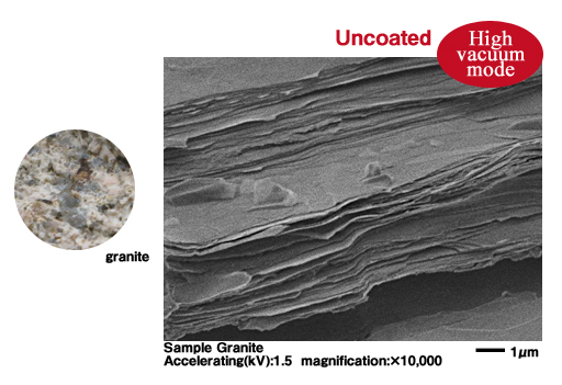
Image Quality
Improved electron optics for higher image quality, faster imaging and analysis.
Less Charging
New scan mode inhibits charging artifacts with non-conductive samples.
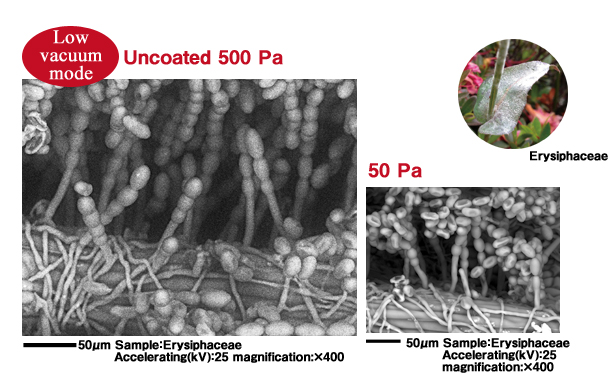
Low Vacuum Mode (LV)
Low vacuum pressure range from 10 to 650Pa extends the range of materials that can be readily observed.
Specimen Navigation
Fully automated, 5 axis motor stage speeds up search for region of interest (ROI).
Quickly reach the ROI from a color image using the Stage Navigation System (Option).
Analysis
High Current mode, probe current up to 1μA (≥20kV).
Advanced Zoom Condenser Lens minimizes image and focus shift due to changes in probe current.
Computer eucentric stage rotation allows for easy positioning of a specimen or ROI.
Versatile
Large Chamber and Stage
Can accept large and heavy specimens, from 200mm diameter × 80mm tall and up to 2kg.
Analytical Port Geometry
Multiple ports for a variety of accessories such as: EDS, EBSD and WDS. Co-planar EDS and EBSD geometry. Dual
EDS detectors can be mounted at 180° for high throughput microanalysis.
Faster Stage Movement
Newly developed motorized stage enables faster positioning (1.5 times faster, compared to other JEOL products).
Fully Automated 5 Axis Motor Stage
Mechanically eucentric, asynchronous, 5 axis (X, Y, Z, T, R) motor control stage.
Computer eucentric tilt and rotation correction built in for tall specimens or when the ROI is not centered on the rotation axis.
New Vacuum System
New vacuum system for fast pump down after specimen exchange. Better signal in low vacuum mode.
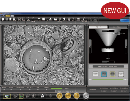
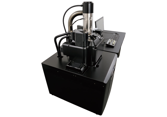
Intuitive Operation
In Touch Interface
Comfortable operation is achieved with a newly designed and developed graphical user interface (GUI), which is operate through the touch screen. The GUI size is adjustable so that the work area can be optimized while analysis or data processing is performed.
Multi-user/Multi-task
Multiple user login function for customized workspace and specimen condition settings.
Multi-language
Interchangeable English to Japanese GUI (select through Icon).
Specifications
| Resolution HV mode | 3.0 nm (30 kV) 15.0 nm (1.0 kV) |
|---|---|
| Resolution LV mode | 4.0 nm (30 kV BED) |
| Magnification | ×5 to ×300,000 |
| Preset magnifications | 6 levels (Can be set by the user) |
| Electron gun | Fully automatic/Manual adjustment available |
| Filament | Factory pre-centered tungsten hairpin filament |
| Accelerating voltage | 0.3 kV to 30 kV |
| Probe current | 1 pA to 1 μA |
| Low vacuum pressure setting range*1 | 10 to 650 Pa |
| Condenser lens | High precision zoom condenser lens |
| Objective lens | Conical lens |
| Objective-lens aperture | 3-step selectable with click-stops
Fine position adjustment in X and Y possible. |
| Astigmatism memory | Available |
| Image shift | ±50 μm (WD 10 mm) |
| Automatic function | Focus
Brightness/Contrast, astigmatism correction |
| Specimen exchange | Changed by opening the specimen chamber door |
| Maximum specimen | Diameter: 200 mm Height: 80 mm (WD10 mm) |
| Specimen stage | Eucentric goniometer stage
(5 axes motor drive stage) X:125 mm Y:100 mm Z:80 mm Tilt: -10 to 90° Rotation: 360° |
| Recipe | Available |
| Image mode | Secondary electron image, REF image,
Composition image, Topographic image, Shadow image |
| Comparison window (Snap shot) | 6 shots (Save, load, imaging condition can be reproduced) |
| OS | Windows®7 |
| Monitor | 23-inch touch panel
(Display resolution 1,920 × 1,080) |
| Number of pixels | 640 × 480 1,280 × 960
2,560 × 1,920 5,120 × 3,840 |
| Image display mode | Multi view, Full screen view, Flexible view,
Add Signals |
| Look up table | Available (Gamma correction, pseudo-color) |
| Histogram display | Available |
| Measurement function | Available (Distance between 2 points,
distance between parallel lines, diameter, etc.) |
| 3D measurement | Available*2 |
| Anaglyph image | Available |
| Report creation software | Built in |
| Language configuration | Available on UI (Japanese/English) |
| Image format | BMP,TIFF,JPEG |
| Image auto save | Available |
| Evacuation system | Fully automatic system
TMP: 1 RP: 1 or 2 *1 |
| Switching vacuum mode | Available on UI (HV/LV) |
Windows is a registered trademark of Microsoft Corporation in the United States and other countries.
Main options
Backscattered electron detector*1
Low vacuum secondary electron detector
Energy dispersive X-ray analyzer (EDS)
Wave length dispersive X-ray analyzer (WDS)
Crystal Orientation Analyzer
Load lock chamber (Preliminary exchange chamber)
Stage navigation system
Chamber scope
Operation panel
LaB6 electron gun
3Dmeasurement software*2
Operation table
Standard feature only for LV/LA
Dedicated software is required for measurement
Specification subject to change without notice.
Application
Application JSM-IT300
Gallery
More Info
Are you a medical professional or personnel engaged in medical care?
No
Please be reminded that these pages are not intended to provide the general public with information about the products.