Aqua Cover method
Aqua Cover method
A method to observe water using a scanning electron microscope (SEM) by keeping water at the liquid state. The method is called the Aqua Cover method because a paper (aqua cover) containing water is used to cover a specimen to prevent evaporation of water in a specimen. When the specimen temperature, the pressure of the specimen chamber and the partial pressure of water vapor are properly controlled, water, aqueous solution and water contained in the specimen can be kept in a liquid state from inserting the specimen into the SEM to observing. The method enables us to observe SEM images of water and the water evaporation process, and of specimens still containing water at higher resolution than optical methods (optical microscope).
Since ordinary SEM observation is performed in a vacuum (<10-2 Pa: atmospheric pressure: 1013 hPa), water is evaporated and thus impossible to observe. However, combination of a low vacuum and a Peltier-cooling stage achieves a liquid state of water (indicated by green area in Fig. 1) in the specimen chamber. Actually, the specimen temperature and the specimen-chamber pressure are respectively set to approximately 0 °C and 650 Pa.
Water is evaporated during evacuation of the chamber from atmospheric pressure to 650 Pa. To avoid evaporation, the specimen is covered with an aqua cover to keep the pressure inside the cover at a saturated water vapor pressure.
Fig. 2 shows the procedures from specimen setting to observation using the method [1]. A Peltier-cooling stage is mounted on the SEM specimen stage and a specimen is placed on the cooling stage and covered with an aqua cover while the specimen temperature is kept at 0 °C by cooling (a). After the specimen chamber is evacuated, the aqua cover is captured by a specimen exchange rod incorporated in the pre-evacuation chamber and then, the cooling stage is lowered (b). The aqua cover is retracted into the pre-evacuation chamber (c). The cooling stage is elevated and SEM observation is performed (d). It is necessary to quickly observe the specimen because water is gradually evaporated after the removal of the aqua cover.
Fig. 3 shows observation examples of liquid droplets leaked from leaf veins using the Aqua Cover method. A movie shows evaporation of water droplets on fibers.
The method enables us to successfully observe fine movements of water droplets during water repellency, which are adhered to the surface of leaves with a micrometer-order structure [2]. This method has also succeeded in detailed observations of water-rich specimens such as organisms and plants, and observations of changes of thickness of fibers and gaps between fibers during swelling and drying processes [3].
There is a method to observe a specimen at its liquid state without using the aqua cover method, in which a specimen is frozen before setting it in the specimen chamber and it is melted after vacuum evacuation for subsequent observation. However, when the specimen is frozen, it can be damaged due to ice-crystal growth or volume expansion. The Aqua Cover method has an advantage that specimen freezing in advance of specimen setting is not required.
This unique method, which enables SEM observation while keeping water in a specimen, is developed by JEOL for the first time. The method is also called the Wet Cover method in the published papers.
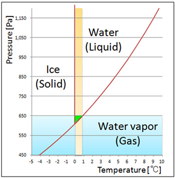
Fig. 1 Schematic of the state diagram of water.
When water is cooled to about 0 °C and the atmospheric pressure is set to approximately 650 Pa, water is preserved at the liquid state.
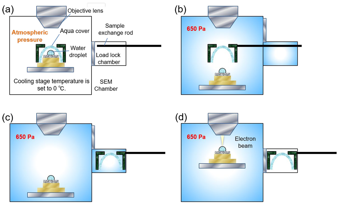
Fig. 2 Observation procedures using the Aqua Cover method.
(a) A Peltier-cooling stage is mounted on the SEM specimen stage at atmospheric pressure. A specimen is placed on the cooling stage and is kept at about 0 °C. Then, the specimen is covered by an aqua cover and the specimen chamber is evacuated until the pressure reaches the set pressure of 650 Pa.
(b) After the pressure reaches 650 Pa and becomes stable, the aqua cover is captured by a specimen exchange rod incorporated in the pre-evacuation chamber. Then, the cooling stage is lowered.
(c) Aqua cover is retracted into the pre-evacuation chamber.
(d) The cooling stage is elevated and SEM observation is performed.
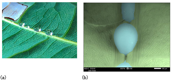
Fig. 3 Liquid droplets leaked from leaf veins at the back of leaves of Pachira aquatica.
(a) Optical microscope image
(b) Backscattered electron image (liquid droplets and leaves are respectively colored by blue and green).
Accelerating voltage: 25 kV, Specimen chamber pressure: 650 Pa.
The water evaporation video (10x speed)
◆Click the "replay" button in the box above, and the movie will start (for 15 seconds)◆
References
[1] N. Inoue, Y. Takashima, M. Suga, T. Suzuki, Y. Nemoto, O. Takai, Observation of wet specimens sensitive to evaporation using scanning electron microscopy, Microscopy, 67 (2018) 356.
[2] N. Inoue, T. Suzuki, Y. Takashima, S. Asahina, H. Onodera, O. Takai, Study of new method for water repellent treatment using scanning electron microscope, Surf. Finish. Soc. Jpn., 70 (2019) 157.
[3] N. Inoue, T. Suzuki, Y. Takashima, M. Suga, O. Takai, Dynamic observation of liquid retained from atmospheric pressure by Wet Cover method using scanning electron microscope, Surf. Finish. Soc. Jpn., 70 (2019) 45.
Term(s) with "Aqua Cover method" in the description
Are you a medical professional or personnel engaged in medical care?
No
Please be reminded that these pages are not intended to provide the general public with information about the products.


