Lorentz electron microscopy
Lorentz electron microscopy
A method to observe magnetic domain structures of a ferromagnetic material using a transmission electron microscope (TEM). Electrons incident onto a ferromagnetic material undergo a force perpendicular to the magnetization direction of the ferromagnetic specimen (Lorentz force), thus their traveling direction changes, as shown in Fig. 1 (electrons are deflected). Since electrons passing through the adjacent domains of the specimen are deflected in opposite directions, this phenomenon enables us to study magnetic domain structures of the specimen. “Lorentz electron microscopy” has two modes which are widely used: Fresnel mode and Foucault mode.
In the Fresnel mode, as shown in Fig. 2, in the adjacent magnetic domains with magnetization direction being different by 180° to each other, the incident electron beams are deflected in the opposite directions. At the left magnetic domain wall, the electron beams passing through the adjacent magnetic domains are deflected closer (overlap) to each other to increase the intensity of the electron beams (Fig. 2(a) to (c)). To the contrary, at the right domain wall, the electron beams passing through the adjacent domains are deflected away (open) to each other to decrease the intensity of the electron beams (Fig. 2(a) to (c)).
Since the overlapped (open) area of the electron-beams is narrow at the bottom surface of the specimen, the magnetic domain wall is difficult to observe when the lens is focused on the bottom surface. When the focus is displaced (defocus), the overlapped (open) area is enlarged and the magnetic domain wall is clearly observed as light (dark) lines (Fig. 2(d) to (f)). The Fresnel mode is also called “defocus method” because it uses the defocus technique.
In the Foucault mode, as shown in Fig. 3, the diffraction spots from the adjacent magnetic domains with magnetization direction being different by 180° to each other, are formed at slightly different positions on the back focal plane, and one of these spots is selected for imaging. The domain that corresponds to the selected diffraction spot appears light whereas the domain corresponding to the unselected diffraction spot appears dark. Since the Foucault mode does not need the defocus technique, this mode is also called “infocus method”.
In addition to these two modes, “electron holography” and “differential phase contrast (DPC)” technique are used.
In a usual TEM, since the specimen is placed in a strong magnetic-filed (maximum~2T) of the objective lens, the specimen is magnetized in the direction of the incident electron beam (magnetic field) and the entire specimen becomes a single magnetic domain. Thus, to observe the magnetic domain structure, it is necessary to turn off the objective lens and to use another lens in the imaging lens system (objective mini lens) under a condition where the specimen is not subjected to a magnetic field.
It is noted that TEMs equipped with a special objective lens applying nearly no magnetic field to the specimen position have also been developed.
Fig. 4 and 5 show usual and Lorentz TEM images of the magnetic domain structure of a permanent magnet of Nd2Fe14B, observed from different directions.
Fig. 4(a), (b) and (c) show Lorentz TEM images taken by the Fresnel mode. In Fig. 4, the magnetization direction of the specimen is approximately parallel to the incident electron beam (perpendicular to the specimen plane), but not completely parallel to the electron-beam direction. As a result, the magnetization components perpendicular to the incident electron beam deflect the beam and provide light and dark magnetic domain walls in a maze-like magnetic pattern. Fig. 4(a), (b) and (c) show respectively the case of infocus, underfocus and overfocus.
Fig. 4(d) and (e) show the enlarged images of the rectangular area of Fig. 4(b) and (c), respectively. The light line and dark line indicated by white and black arrowheads correspond to the magnetic domain wall. In the infocus image (a), the wall cannot be observed because the width of the wall image is too narrow, but in the defocus images (d) and (e), the magnetic domain walls are clearly observed. It is noticed that light and dark images of the magnetic domain wall are reversed by the positive or negative defocus (overfocus or underfocus).
Fig. 5(a) is a usual TEM image and Fig. 5(b) and (c) are Lorentz TEM images taken by the Foucault mode. In this case, the magnetization direction of the specimen is perpendicular to the incidence electron beam (parallel to the specimen plane), and the magnetization directions of the adjacent domains are opposite. In the usual bright-field imaging condition (a), no contrast is observed between the magnetic domains. However, when the aperture position is changed to select one of diffraction spots produced by either of magnetic domains, stripe-shaped magnetic domains are clearly observed (b). When the aperture position is changed to select the other one of diffraction spots, the light and dark images of the adjacent domains are reversed (c).

Fig. 1. Schematic of the deflection of the electron beam due to magnetization B of a magnetic material.
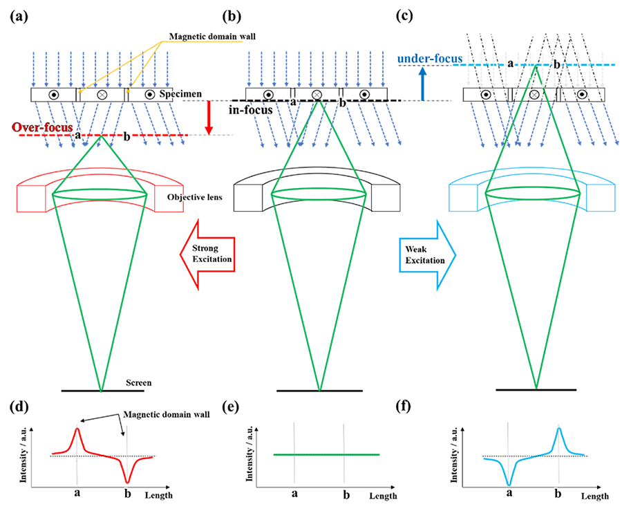
Fig. 2: Schematic of the Fresnel mode.
By increasing or decreasing the lens intensity of the imaging system, the position of the acquired image is changed. (a) overfocus: the lens is adjusted to focus on a plane lower than the bottom surface of the specimen. (b) infocus: the lens is focused on the bottom surface of the specimen, and (c) underfocus: the lens is focused on a plane upper than the bottom surface of the specimen.
The intensity profiles of the magnetic domain wall images in the cases of overfocus, infocus, and underfocus are respectively shown in (d), (e), and (f). In infocus (e), no change in the intensity profile occurs at the magnetic domain walls, but in overfocus (d) and underfocus (f), the magnetic domain walls appear as light (with intensity increase) or dark (with intensity decrease) for the magnetic domain wall “a”. The reversal of image profiles between the underfocus and overfocus cases can be used to determine the direction of magnetization of the specimen.
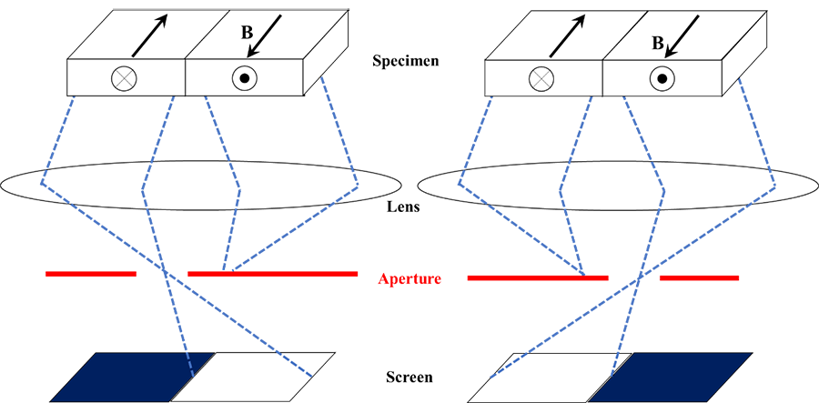
Fig. 3. Schematic of the Foucault mode.
The directions of the deflection of the electron beams are reversed due to the inversion of magnetization directions of the specimen. Diffraction spots are produced at the different positions on the back focal plane of the objective lens (or the objective mini lens). When selecting one diffraction spot by the aperture, the domain that corresponds to the selected diffraction spot appears light whereas the domain corresponding to the diffraction spot unselected by the aperture becomes dark, which makes it possible to visualize the domains.
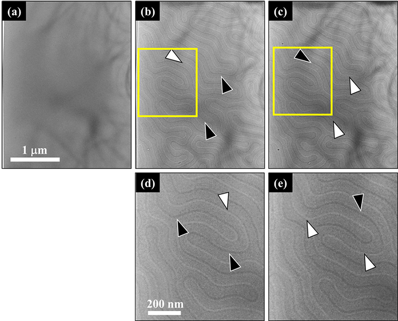
Fig. 4. TEM images of a permanent magnet of Nd2Fe14B, taken by the Fresnel mode at (a) infocus, (b) underfocus, (c) overfocus. (d) and (e) are the enlarged images that correspond to the rectangular area of (b) and (c) respectively. Magnetic domain walls are observed as light- and dark-curved lines, indicated by white and black arrowheads.
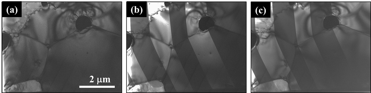
Fig. 5. TEM images of a permanent magnet of Nd2Fe14B, taken by the Foucault mode.
(a) Selecting two diffraction spots produced by both magnetic domains in a usual way. The magnetic domains are not visualized.(b) and (c) Selecting only one diffraction spot produced by either of magnetic domains. One type of the magnetic domain is seen light whereas the other magnetic domain loses intensity. The magnetic domain structure is visualized.
Related Term(s)
Term(s) with "Lorentz electron microscopy" in the description

Are you a medical professional or personnel engaged in medical care?
No
Please be reminded that these pages are not intended to provide the general public with information about the products.


