Mass image quality improvement using a machine learning model "FINE-AI Filter"
MSTips No. 526
Mass imaging (MSI) using matrix-assisted laser desorption/ionization (MALDI) have seen increasing use in various fields in recent years. The soft ionization method of MALDI makes it possible to visualize the localization of various molecules. However, MALDI-MSI suffers from a low signal-to-noise ratio in extracted mass images, mainly for the following two reasons: I) Non-uniformity occurs in the crystal morphology of the matrix sprayed onto the sample surface. II) Each pixel is a local analysis of approximately several tens of micrometers square, resulting in a small number of ions obtained from that region. These issues are particularly pronounced in extracted mass images of trace components with low peak intensity. Therefore, in this report, we attempted to improve the image quality of extracted mass images using the FINE-AI Filter, which was developed by applying a machine-learning model for noise filtering—originally created from secondary electron images obtained by scanning electron microscopy (SEM)—to MALDI-MSI.
Experiment
A machine learning model using secondary electron images from a SEM was created using the following procedure. A supervised machine learning method, conditional generative adversarial network (cGAN), was used for machine learning. First, approximately 3000 high-quality secondary electron images were prepared as training data. Next, low-quality images were created by adding shot noise following a Poisson distribution to these high-quality images at several different signal-to-noise ratio settings. When a low-quality secondary electron image is input into the Generator shown in Figure 1, a high-quality secondary electron image is generated. Training was performed repeatedly by comparing the measured high-quality secondary electron image with the high-quality image generated by the machine-learning model.
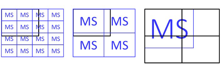
Although the trained Generator was used to improve extracted mass images, the field of view in SEM is fixed whereas the field of view in MALDI-MSI can be set arbitrarily, so the FINE-AI Filter employs several conversion strategies. The image-size adjustment methods are as follows: (i) When the mass image is smaller than the SEM image size, multiple copies are arranged, converted, and then part of the result is cropped out (Figure 2, left). (ii) As a variation of method (i), the mass image is enlarged relative to its acquired size by selecting x1, x2, or x3 magnification before image-quality improvement is performed (Figure 2, center). (iii) When the mass image is larger than the SEM image size, it is divided into multiple sections, converted separately, and then recombined (Figure 2, right).
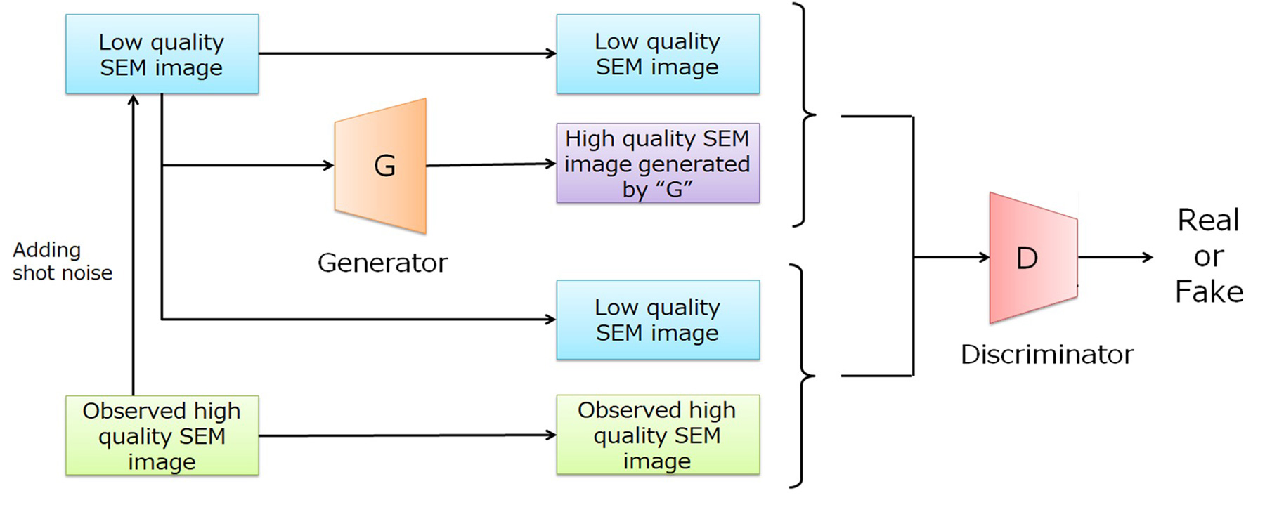
Figure 1 Development of an image quality improvement model using SEM images
The black frame indicates the SEM image size (fixed), and the blue frame indicates the MSI image size (variable).
Figure 2 Image size adjustment when applying machine learning models created from SEM images to mass images.
Results
Mouse brain frozen tissue sections were used as samples, and DHB was sprayed as the matrix. Measurements were performed using the SpiralTOF positive ion mode of a NewSpiralTOF™. The pixel size was 40µm, and the image size was 125×169 pixels. Figure 3 shows the average mass spectrum in the upper left. Various lipids (mainly phosphatidylcholine, phosphatidylethanolamine, and galactosylceramide) were observed between m/z 700-1000. The upper right shows an enlarged mass spectrum around m/z 820. Using NewSpiralTOF™ enabled isobar separation, which is difficult with conventional reflectron-type TOFMS. Many of the observed lipid ions had relatively low ion intensity, resulting in coarse image quality. Therefore, we compared image improvement methods using a FINE-AI filter, a Gaussian filter, and binning. The FINE-AI filter improved image quality while maintaining contours compared to commonly used Gaussian filters and binning. When the magnification of the FINE-AI Filter was changed, it was found that fine details were lost at lower magnifications, while noise reduction was insufficient at higher magnifications. For the results of this measurement, x2 appears to be appropriate. The magnification should be selected according to the sample structure and pixel size.
Conclusion
To improve the image quality of mass images, we attempted to apply a machine learning model for noise filtering, created using a scanning electron microscope. We also investigated processing methods for applying this model to mass images. This method was found to improve the image quality of mass images with low S/N ratios, such as images of very small peaks or images strongly affected by the matrix.
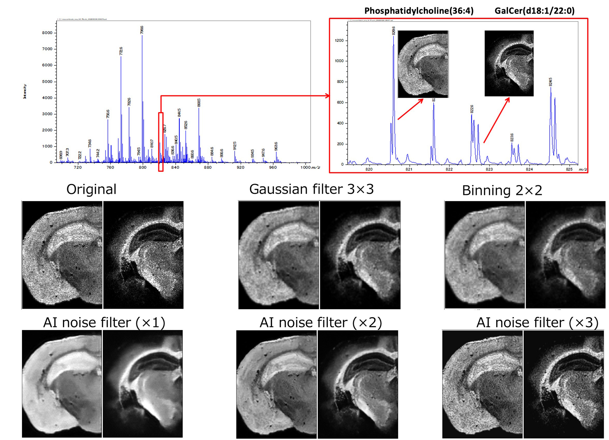
Figure 3 Extracted mass image quality improvement using FINE-AI Filter
Solutions by field
Electrical / Electronic Component
Related products
Are you a medical professional or personnel engaged in medical care?
No
Please be reminded that these pages are not intended to provide the general public with information about the products.