Compositional and Structural Analysis of Labeled N‑Linked Glycans Using JMS‑S3000 NewSpiralTOF™
MSTips No. 519
Introduction
N‑linked glycans are post‑translational modifications in which N‑acetylglucosamine (GlcNAc) is attached to asparagine residues of proteins. They play important roles in protein structure and function and are involved in biological processes such as signal transduction and immune responses. Structural changes in N‑linked glycans are closely associated with various diseases, and they have attracted attention as biomarkers, particularly in cancer and congenital disorders of glycosylation. Therefore, accurate structural analysis of N‑linked glycans is important for both basic research and clinical applications. In this application note, labeled N‑linked glycans (Fig. 1) were analyzed using the JMS‑S3000, and results of accurate mass measurements in the SpiralTOF mode and structural analysis in the TOF‑TOF mode are presented.
Method
2,5‑Dihydroxybenzoic acid (DHB; FUJIFILM Wako Pure Chemical Corporation) was used as the matrix at 10 mg/mL in a 50% ACN–0.1% TFA aqueous solution. A 50 pmol/µL aqueous solution of a labeled N‑linked glycan, 3‑G1 2AB (Tokyo Chemical Industry Co., Ltd.), was used as the analyte. The sample and matrix solutions (0.5 µL each) were spotted onto a target plate and air‑dried to form co‑crystals. Accurate mass measurements were performed in the SpiralTOF mode, followed by structural analysis in the TOF‑TOF mode (both in positive ion mode). The labeled N‑linked glycan 3‑G1 2AB consists of N‑acetylglucosamine (GlcNAc), mannose (Man), and galactose (Gal).
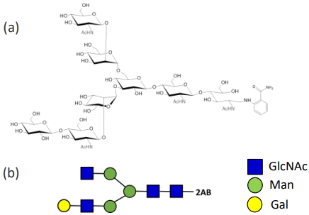
Fig. 1 The structure of Labeled N-glycan 3-G1 2AB(a) and Symbol Nomenclature for Glycans(b)
Result
Measurement result by SpiralTOF mode
The results obtained using the SpiralTOF mode are shown in Fig. 2. The labeled N‑linked glycan 3‑G1 2AB was observed as a sodium adduct ion (Fig. 2‑a). The observed peak exhibited a mass resolving power of over 70,000. Next, accurate mass measurements were performed using angiotensin I (AngI) and adrenocorticotropic hormone fragment 1–17 (human, rat; ACTH 1–17) as internal standards (Fig. 2‑b). The measured mass of the labeled N‑linked glycan 3‑G1 2AB was 1621.5982, showing a mass error of +0.9 mDa relative to the calculated value of 1621.5973. Figure 3 shows the isotope pattern of the sodium adduct ion of the labeled N‑linked glycan 3‑G1 2AB. The observed isotope pattern closely matched the theoretical isotope distribution.
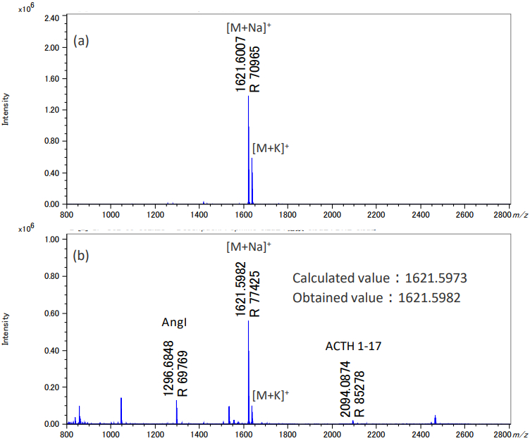
Fig. 2 Mass spectra of Labeled N-glycan 3-G1 AB(a) and Labeled N-glycan 3-G1 2AB with peptide mix(b).
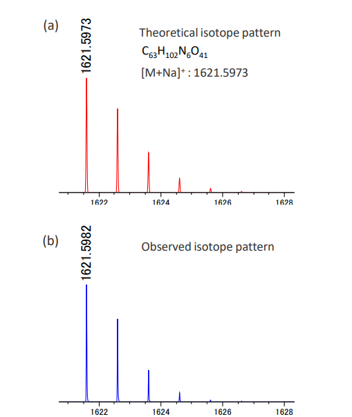
Fig. 3 Comparison of theoretical isotope pattern(a) and obtained isotope pattern(b).
Measurement results using TOF-TOF mode
The product ion spectrum obtained from m/z 1621.6 in the TOF‑TOF mode is shown in Fig. 4. The observation of a peak at m/z 23 confirms that the detected ion of the labeled N‑linked glycan 3‑G1 2AB is indeed a sodium adduct ion. In addition, characteristic X ions (▼) such as m/z 392, 595, and 1122, and Y ions (▼) such as m/z 364, 567, and 1093, in which the charge is retained on the reducing end of the glycan, were clearly observed. X ions are generated by cleavages within the sugar ring and yield characteristic product ions that retain the oxygen atoms contained in the ring structure. In contrast, Y ions are formed by cleavage of glycosidic bonds and are known to reflect the backbone structure of the glycan. From the observed Y ions, product ions derived from GlcNAc (■), mannose (Man, ●), and galactose (Gal, ●) were confirmed. Based on these results, the constituent monosaccharides of the labeled N‑linked glycan 3‑G1 2AB were clearly identified.
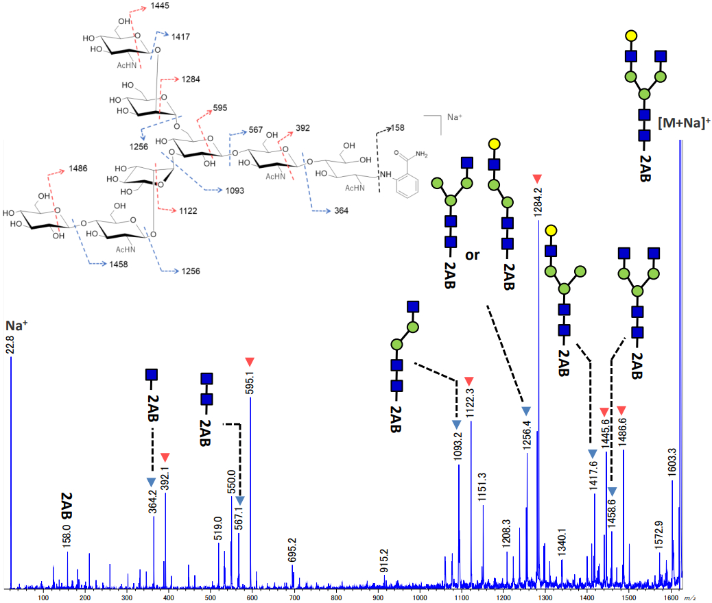
Fig. 4 Product ion spectrum of Labeled N-glycan 3-G1 2AB and proposed assignments
Conclusion
Labeled N‑linked glycans were analyzed by MALDI‑TOFMS. In the SpiralTOF mode, the target peaks were observed with high mass accuracy. In addition, in the TOF‑TOF mode, X and Y ions derived from the glycan ring structure were clearly observed, enabling the identification of the constituent monosaccharides. Based on these results, it was demonstrated that the JMS‑S3000 is capable of both compositional analysis and structural analysis of glycans.
Solutions by field
Related products
Are you a medical professional or personnel engaged in medical care?
No
Please be reminded that these pages are not intended to provide the general public with information about the products.