Mass imaging of strawberry using the JMS-S3000 “SpiralTOF™-plus3.0” and transfer plate Poropare™
MSTips No. 499
Mass spectrometry (MSI) technology using matrix-assisted laser desorption/ionization (MALDI) has been widely used in various fields in recent years. The MALDI method, which is a soft ionization method, can visualize the localization of various molecules. In a typical workflow of MALDI-MSI, a 5-10 μm thick tissue section is prepared and placed on a conductive ITO glass slide. A matrix is uniformly applied on the slice, and MALDI-MSI measurement is performed. One of the challenges of MALDI-MSI is that it is difficult to prepare slices, such as the surface measurement of plants and plastic samples. In particular, fruits and plants contain a lot of water, making it challenging to prepare frozen tissue sections. For such samples, one solution is to transfer the organic compounds on the sample surface to a specially processed plate and measure them. This report used a novel, highly robust transfer plate, Poropare™ (Hamamatsu Photonics), to transfer compounds in strawberries and perform MSI measurement. Poropare™ is made by sintering glass beads of several tens of μm and depositing platinum on them to ensure electrical conductivity (Figure 1). This platinum effect also makes it possible to perform surface-assisted desorption ionization (SALDI-MSI) without a matrix. In this study, we examined both SALDI-MSI and MALDI-MSI. Furthermore, the high-mass resolution MALDI-TOFMS JMS-S3000 "SpiralTOF™-plus 3.0" used in this report employs a SpiralTOF ion optical system consisting of four electric sectors and has a long flight distance of 17 m. This makes it less susceptible to the unevenness of the Poropare™ surface, achieving high resolution. In addition, excluding post-source decay ions by the electric sectors allows for high-sensitivity measurements even in the low molecular weight region.

Figure 1 Schematic of Poropare™
Experiments
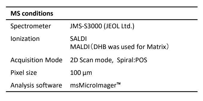
Commercially available strawberries were prepared and cut into four pieces with a knife. One of the pieces was pressed against a Poropare™ to transfer the components present at the cut surface (Figure 2). In addition, the surface of the same strawberry was transferred in the same manner, and then DHB was applied to the matrix with an airbrush. MSI measurements were performed in SpiralTOF positive ion mode using a JMS-S3000 SpiralTOF™-plus 3.0. The obtained data was analyzed using an msMicroImager™. Other detailed measurement conditions are shown in Table 1.
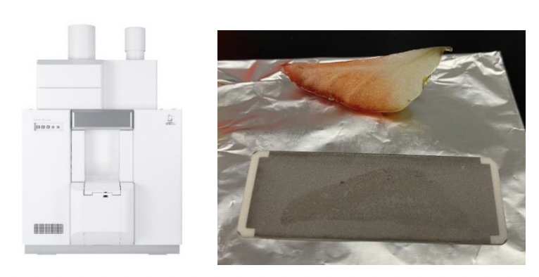
Figure 2 Sample preparation using Poropare™
Table 1 Measurement conditions
Poropare is trademark of Hamamatsu Photonics K.K.
Results
Figure 3 shows the results of SALDI-MSI on a cut strawberry. Sweet components such as hexose and sucrose, and sour components such as citric acid were observed. The mass error of each component was less than a few mDa, confirming good mass accuracy. Next, MALDI-MSI measurements were performed using a sample coated with DHB, and the results are shown in Figure 4. Hexose, sucrose, and citric acid were also observed as major components in MALDI-MSI. On the other hand, MALDI-MSI was able to observe Pelargonidin-3-O-glucoside, which SALDI-MSI could not observe. Although this component was minor, it was separated from another component that differed by about 0.1 Da, and the mass error was -1.6 mDa. This is the result of taking advantage of the high resolution characteristics of the SpiralTOF™-plus 3.0 in the measurement of the low molecular weight region. Pelargonidin-3-O-glucoside is the major anthocyanin pigment in strawberries and was found to be distributed on the outside of the strawberry.
Conclusion
Using a Poropare™, we investigated MSI measurement of components inside strawberries, which are generally challenging to make tissue sections for mass imaging. Although the transfer plate has a large uneven structure due to the transfer function, it was found that by combining it with the SpiralTOF™-plus3.0, which has a long flight distance, mass imaging with high resolution and high mass accuracy is possible even in the low molecular weight region.
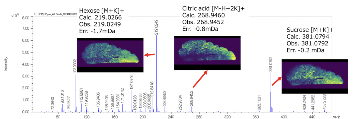
Figure 3 Average mass spectrum of the result of SALDI-MSI and the extracted mass images of Hexose, Sucrose, and Citric acid.
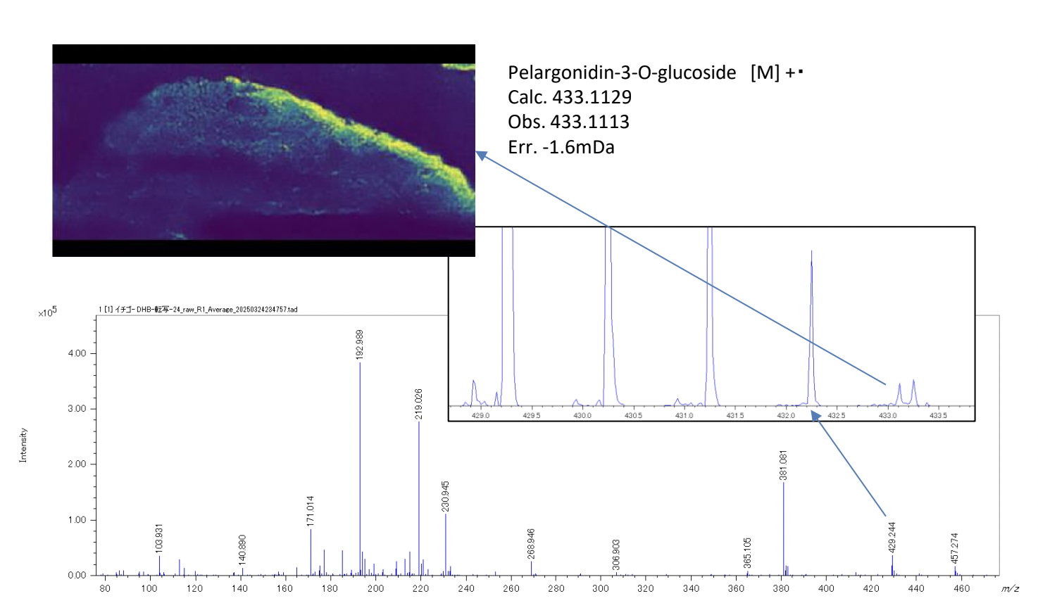
Figure 4 Average mass spectrum of the result of MALDI-MSI and extracted mass image of Pelargonidin-3-O-glucoside.
Solutions by field
Related products
JMS-S3000 SpiralTOF™-plus 3.0Matrix-Assisted Laser Desorption/Ionization Time-of-FlightMass Spectrometer
The JMS-S3000 SpiralTOF™-plus 3.0 is a MALDI-TOFMS* that uses innovative SpiralTOF ion optics that has been updated to extend the mass range of this high resolution system. The JMS-S3000 defines a new standard in MALDI-TOFMS performance and provides state-of-the-art analytical solutions for a wide range of research areas such as functional synthetic polymers, materials science, and biomolecules.Product category
Are you a medical professional or personnel engaged in medical care?
No
Please be reminded that these pages are not intended to provide the general public with information about the products.