High Mass-Resolution MALDl-imaging MS for Drug Metabolism in Tissue Using the JMS-S3000 “SpiralTOF™”
MSTips No. 212
Introduction
Imaging by using matrix-assisted laser desorption/ionization mass spectrometry (MALDI-Imaging) has been expanded during the last decade into biological applications in order to assess the distribution of proteins, peptides, lipids, drugs, and metabolites in tissue specimens. For a drug metabolism analysis, MALDI-Imaging has an advantage in that it can visualize the distributions of drugs and metabolites without using radio isotopes which are used for whole body autoradiography.
In MALDI-Imaging measurements, a laser is used to irradiate each point across a sample surface in order to acquire a mass spectrum for a given location. By combining the mass spectra with their two-dimensional position information, localization of compounds with inherent molecular weights can be visualized or the mass spectra for certain regions of interests (ROIs) can be created.
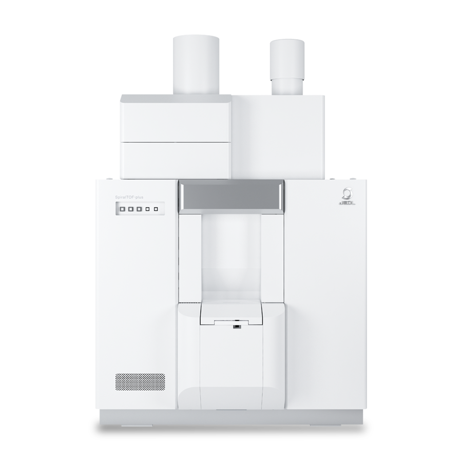
The JMS-S3000 SpiralTOF™ is a MALDI-TOFMS, which utilizes the JEOL patented spiral ion optics system. It has a 5-10 times longer flight path than the typical reflectron type MALDI-TOFMS. As a result, it can achieve high mass-resolution to separate peaks that have the same nominal mass but have different exact masses (isobaric separation). This feature is particularly effective for MALDI-Imaging for drug metabolism, which typically consist of relatively low molecular weight compounds which are often interfered with by matrix compounds and/or surface contaminants.
Experiments
Sample and preparation conditions are listed in Table1.
Table 1 Sample and sample preparation conditions.
Sample and preparation conditions are listed in Table1.
The MS Imaging measurements on the liver tissue section (7.8 mmx9.2 mm) were performed on the SpiralTOF in positive ion mode with a spatial resolution of 50 µm. The MS Imaging data was processed with msMicroImager (JEOL). The MS/MS measurements with TOF-TOF positive ion mode were also performed for structure analysis of the drug and its metabolite.
Results and Discussion
The peak observed at m/z 472.3425 in the averaged mass spectrum was assigned to terfenadine (C32H41NO2) [M+H]+ (m/z 472.3210) and was supported by the MS/MS measurements (described below). The averaged mass spectrum, which was mass corrected using the assigned peak of terfenadine, is shown in Fig. 1. The enlarged mass spectrum at m/z 472 and 502 are also shown. The peak at m/z 502.2944 was assigned to fexofenadine (C32H39NO4) [M+H]+ (m/z 502.2952), a metabolite of terfenadine, by accurate mass and MS/MS measurements (also described below). The isobaric separation capability of 0.2-0.3 u was then used to draw the inherent mass images for each target peak. The optical image of the tissue section and the mass images for m/z 472.3 and m/z 502.3 with 0.1u mass window are shown in Fig.2. Both terfenadine and fexofendine were distributed across the liver tissue sections.
The product ion spectrum of a) terfenadine spotted on ITO glass, b) m/z 472.3 and c) m/z 502.2 from the liver tissue section are shown in Fig. 3. The fragmentation channels observed in Fig. 3a and b were nearly identical so that m/z 472.3 was assigned to terfenadine. The estimated fragmentation paths observed at m/z 216, 270 and 288 are shown in the structural formula for terfenadine. Most of the fragments observed in Fig. 3c were similar to Fig 3a, but several of them were observed to have a 30 u difference (red numbers). These differences were likely due to the methyl group in terfenadine changing to a carboxyl group in fexofendine.
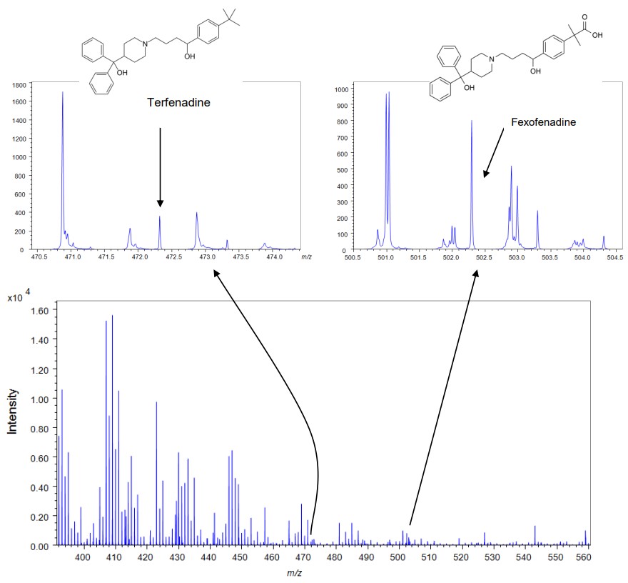
Fig. 1 Averaged mass spectrum of all pixels acquired in IMS measurement.
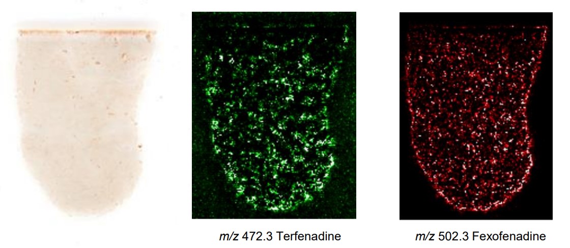
Fig.2 Pictures of tissue section and extracted mass image at m/z 472.3 and 502.3
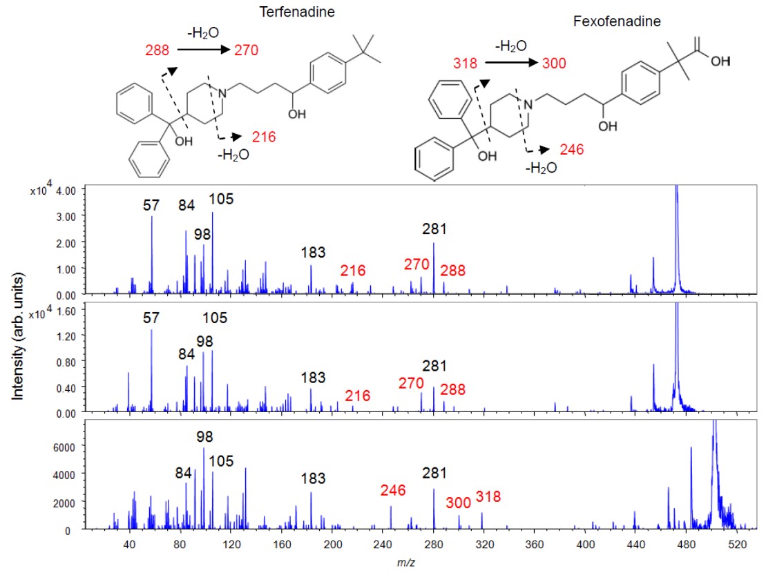
Fig.3 The product ion spectrum of m/z 472.3 from standard terfenadine(a). The product ion spectra from the liver tissue section, m/z 472.3(b) and m/z 502.3 of fexofenadine (c).
Acknowledgment
We would like to thank Daiichi Sankyo Co., Ltd. for providing the mouse liver tissue sections.
Solutions by field
Related products
Product category
Are you a medical professional or personnel engaged in medical care?
No
Please be reminded that these pages are not intended to provide the general public with information about the products.