Observation of amosite asbestos cross section microstructure by a high-resolution scanning transmission electron microscope
EM2025-06
Introduction
Among amphibole asbestos, crocidolite asbestos is considered to be highly toxic to the human body, as it tends to be miniaturized and easily adsorbed. For the miniaturization, many studies have been conducted (i.e. [1]) and indicated that crocidolite asbestos is a gathering of multi-crystal microfibers and that there is a possibility where many improper layering and layered silicate are involved [1]. We conducted an optimal bright field-STEM (OBF-STEM) [2], a kind of scanning transmission electron microscopy (STEM) to observe a cross section of crocidolite asbestos. Due to OBF-STEM, which enables high contrast atomic resolution observation under low electron beam condition, we could elucidate that the layered silicate minerals existing in the gaps of crocidolite asbestos were talc-like silicate [3]. On the other hand, a possibility is also indicated that amosite asbestos, another amphibole asbestos may have a significant influence to the human body, as it has a high aspect ratio [4]. However, there has been no detailed research with detailed cross-sectional observation, and its miniaturization mechanism is unknown. To elucidate the miniaturization mechanism, it is important to bring about the similarity and difference in terms of mineralogy with other asbestos. In this study, we observed miniaturized tissue of amosite asbestos by using high -resolution STEM, in order to compare with crocidolite asbestos.
1. Experiment condition
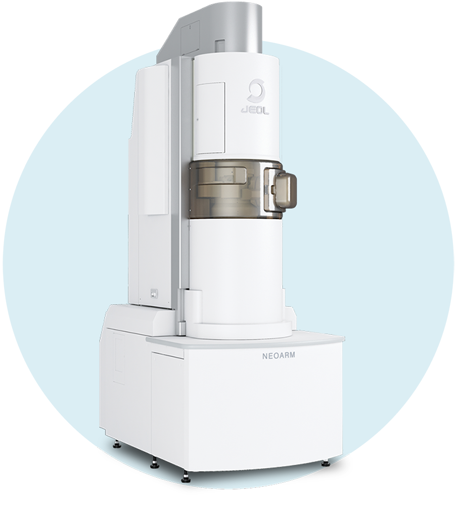
JEM-ARM200F
- Instrument:JEM-ARM200F NEOARM (HR)
- Acceleration Voltage :200 kV
- Probe current:~10 pA
- Specimen:Amosite Asbestos from South Africa
Crocidolite from Cape (for comparison)
2. Comparison of amosite and crocidolite
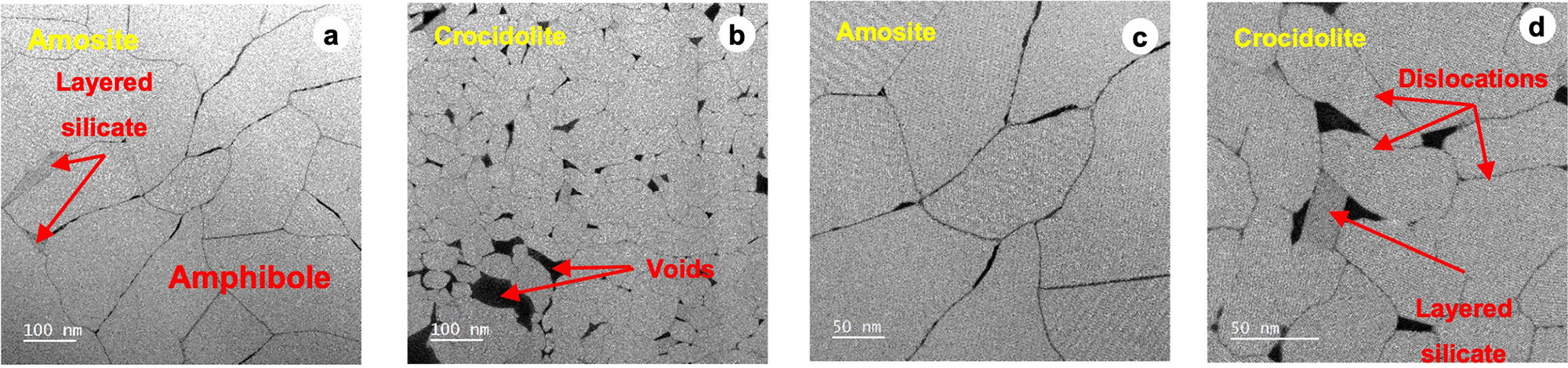
Fig. 1 ADF-STEM low magnification image (a)(b) and middle magnification image (c)(d) of amosite & crocidolite
The microfiber size is different between amosite and crocidolite asbestos. The size of the voids between microfibers is also different (a)(b). With amosite, layered silicate often exists as if filling in the gaps, such as the grain boundary triple point of microfibers (a). With crocidolite asbestos, dislocations are seen in the microfibers (d), while dislocations are rarely seen in the microfibers of amosite asbestos (c).
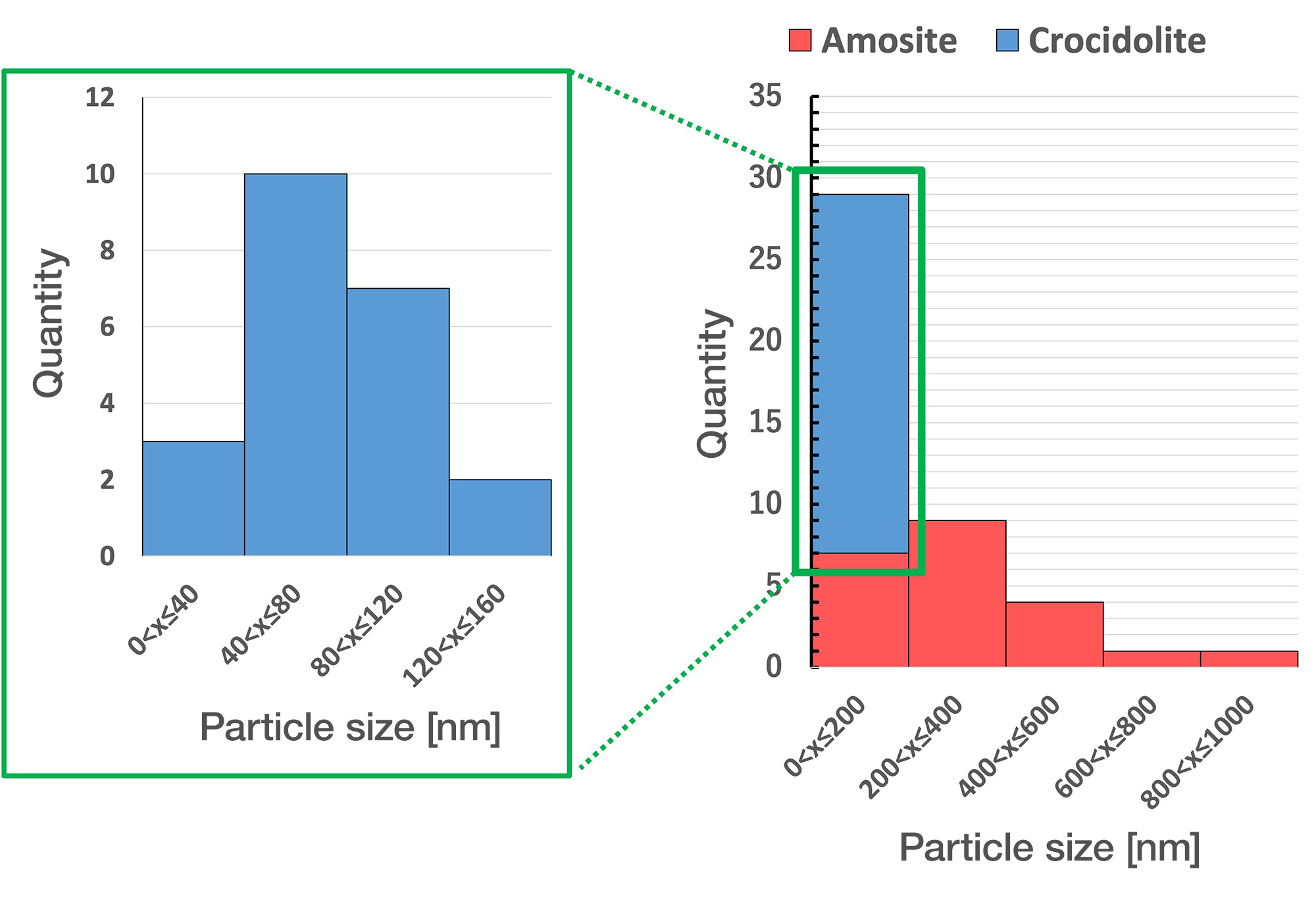
Fig. 2 Particle size distribution of amosite & crocidolite
Long diameter of amosite microfiber is approximately 100 to 1000nm, the most frequent value is "200<x≦400" rank. The long diameter of crocidolite is approximately 10 to 200nm, the most frequent value is "40<x≦80" rank.

Fig.3. ADM-STEM middle magnification image (a)(b) and atomic resolution image (c)(d) of layered silicate and dislocation of amosite
In amosite, there is hardly any dislocation seen in the microfiber (Fig.1). However, dislocations exist limitedly in microfibers around the layered silicate filling in the grain boundary(a)(b). Dislocations consist of a triple chain (c). There are areas where the boundaries of triple chains are connected by a multi-chain silicate (d).
3. Layered silicate in amosite
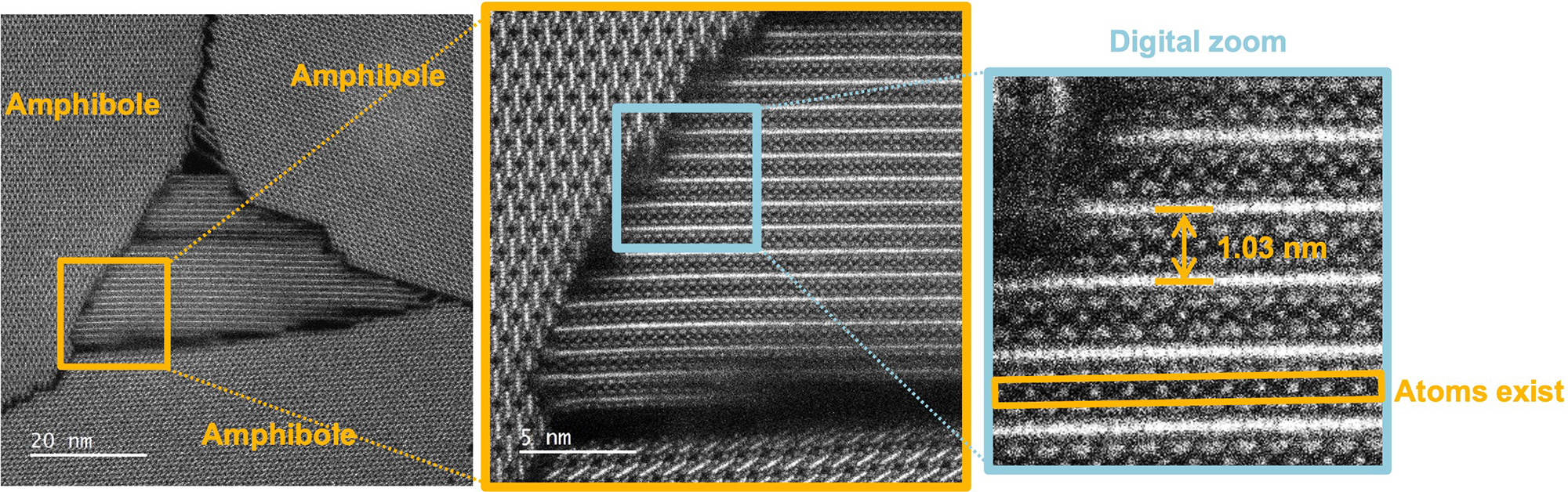
Fig. 4. ADF-STEM atomic resolution image of a layered silicate in amosite
In the layered silicate filling in the grain boundary in amosite, interlayer atoms exist, and the baseline spacing (d002) of the layer is up to 1.03 nm. Therefore, it is indicated that the layered silicate mineral in amosite is a mica-like silicate mineral.
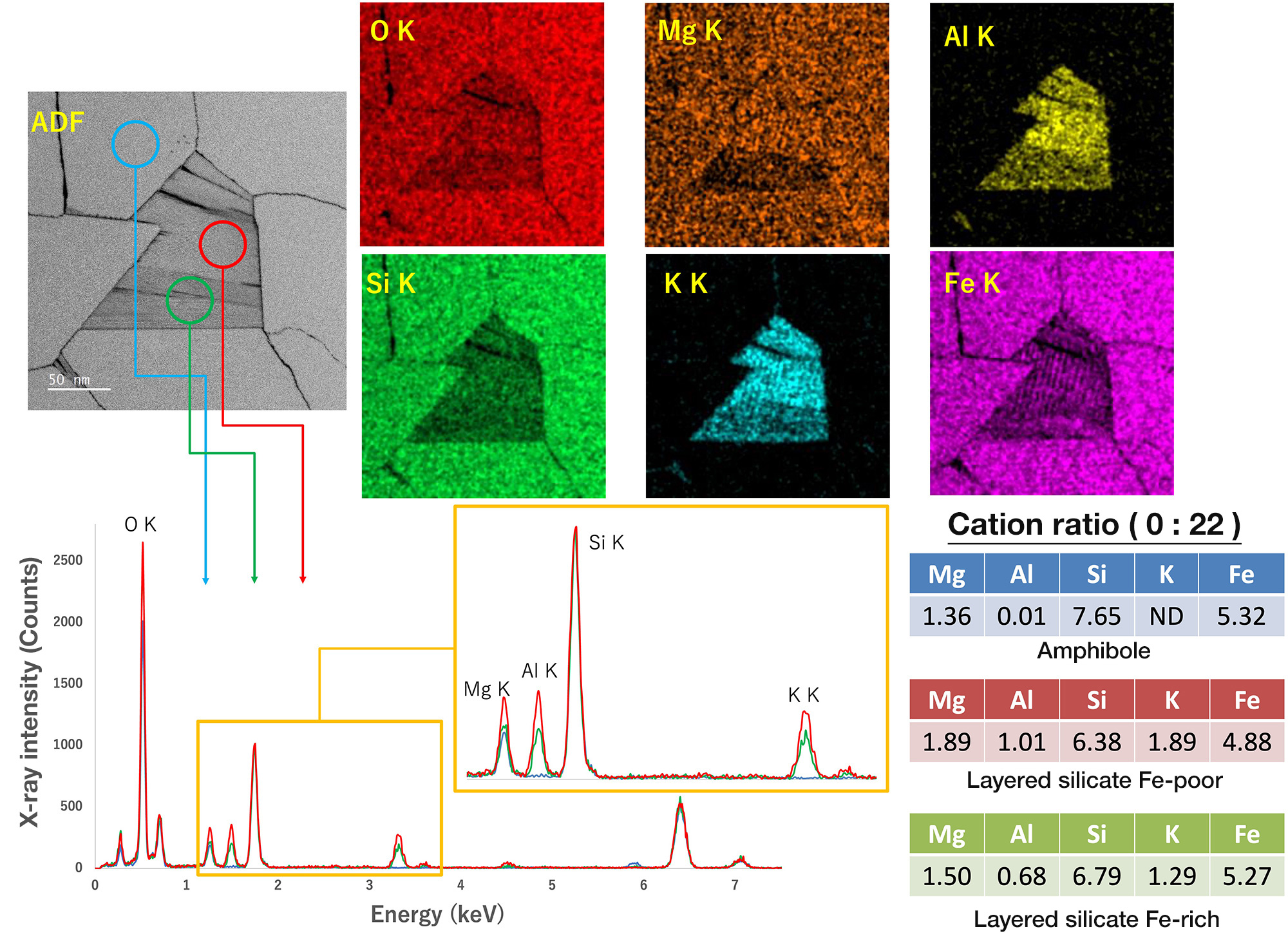
Fig.5. Extracted spectra from EDS elemental map & elemental map of amosite (Si-normalized)
Layered silicate in amosite is rich with elements such as potassium, magnesium, and iron. Based on the cation ratio, it is considered a layered silicate that is close to mica. In particular, for iron, magnesium, aluminum, the difference of concentration distribution is seen according to the location.
As a result of ADF-STEM observation and EDS analysis, it is revealed that
mica or mica-like silicate mineral exists in amosite.
Summary
Microstructure of amosite asbestos was observed by using a high-resolution STEM.
By comparing with crocidolite, there is a difference of long diameter of microfiber, distribution of multiple chain silicate & improper layering.
As a result of ADF-STEM observation, it is revealed that mica or mica-like silicate exists in amosite.
EDS analysis revealed that layered silicate has compositional diversity
Reference
[1] J.H. Ahn & P.R. Buseck (1991) Am. Min., 76, 1467-1478.
[2] Ooe et al. (2021) Ultramicroscopy 220, 113133.
[3] Onishi & Miura. (2023) *Nihon Koubutsu Kagakukai 2023nen nenkai kouen yousishu”[Japan Association of Mineralogical Sciences 2023 Annual Meeting Seminar Abstracts] 279-280.
[4] Murai.(2006), *Ganseki Koubutsu Kagaku” [Journal of Mineralogical and Petrological Science]35, 34-39
Solutions by field
Related products
Are you a medical professional or personnel engaged in medical care?
No
Please be reminded that these pages are not intended to provide the general public with information about the products.