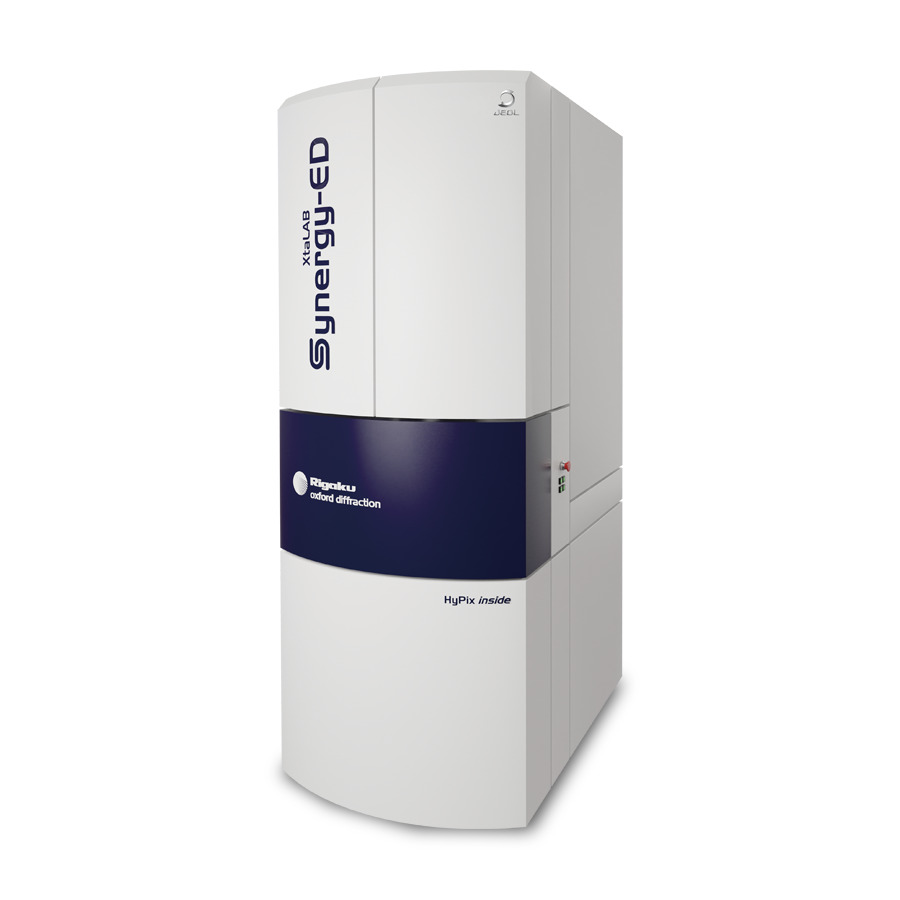
Single Crystal Analysis by XtaLAB Synergy-ED
ED2022-01E
A novel method for determining the structure of submicron particles
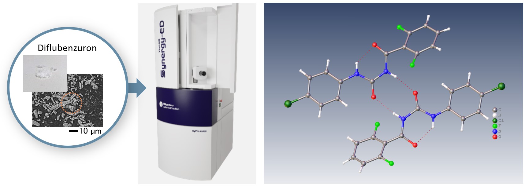
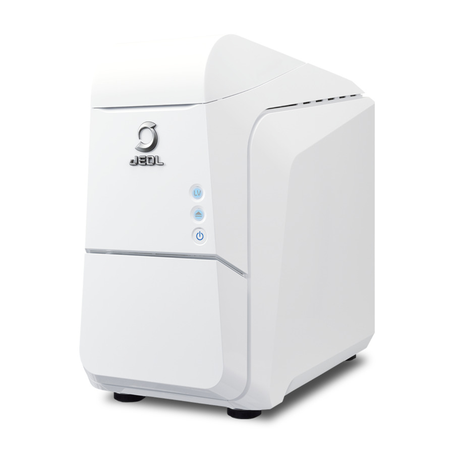
Left : Backscattered electron image of Diflubenzuron in LV-mode, JCM-7000 NeoScope™
Right : Electron diffraction structure analysis of Diflubenzuron, XtaLAB Synergy-ED
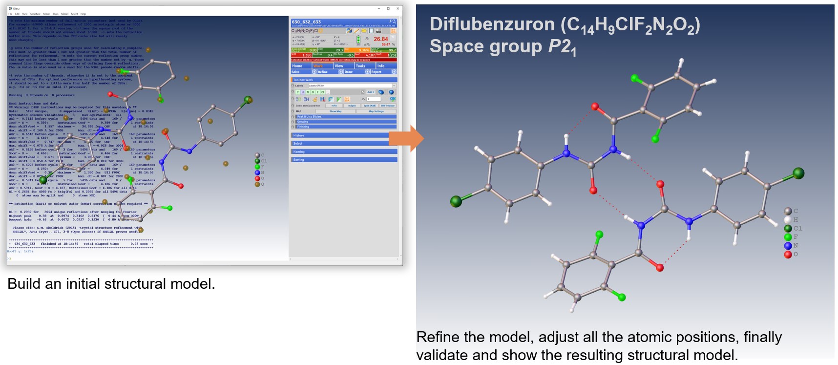
XtaLAB Synergy-ED is a fully optimized electron diffractometer for microcrystals analysis, jointly developed by both companies: Rigaku Corporation and JEOL Ltd. The XtaLAB Synergy-ED features Rigaku’s high-speed and high-sensitivity photon-counting detector (HyPix-ED) and a state-of-the-art instrument control with a single crystal analysis software platform (CrysAlisPro for ED). The key characteristic of this product provides researchers an easy and efficient platform for electron crystallography. In the example above, the right-hand shows the result of structural analysis of Diflubenzuron (C14H9ClF2N2O2).
Sample preparation
No sample preparation is required: load your sample directly onto a TEM grid as follows. If your particles are large, grind them with a microscope slide or spatula to a fine particle size.
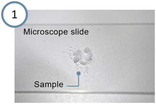
Place your sample on the microscope slide.
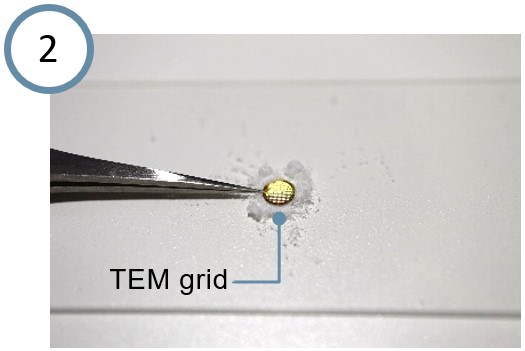
Attach your sample directly to the TEM grid and brush off excess particles.
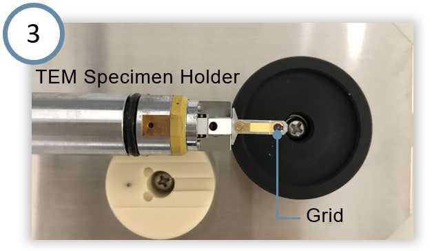
Insert the grid into the specimen holder.
3D ED/microED experiments
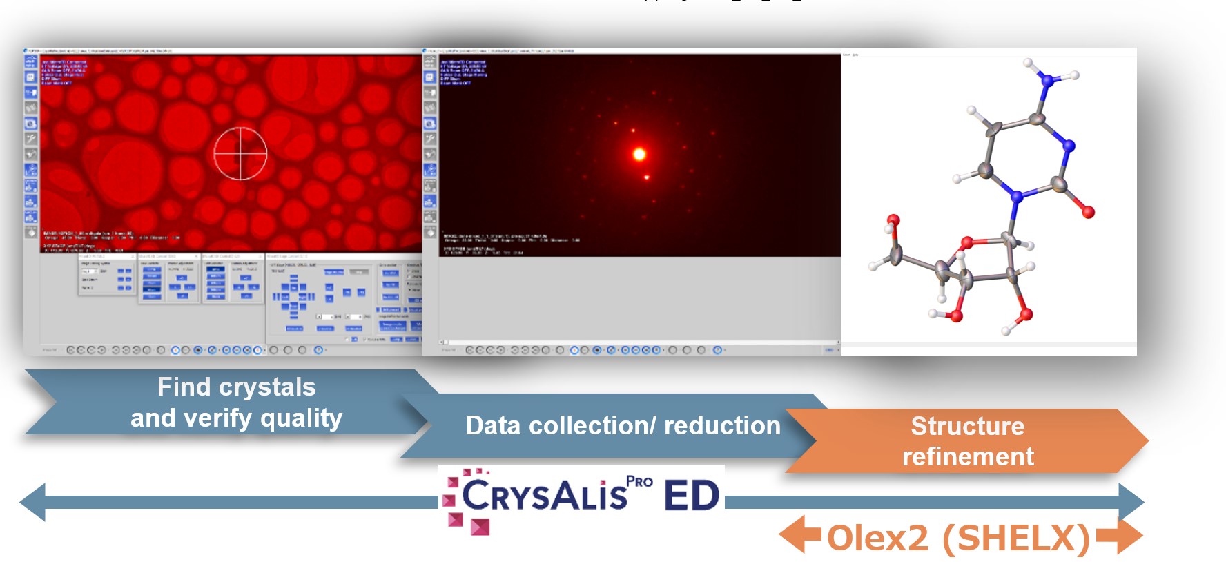
CrysAlisPro for ED allows easy switching between the visual mode and the diffraction mode. In the visual mode, click on an appropriate particle and the Diff.exp button in the MicroED Stage Control menu to switch into the diffraction mode. And then click the Start experiment button in the MicroED/3D ED DC window to start the electron diffraction observation automatically. The rotation range used for a typical measurement is −60 to +60 degrees.
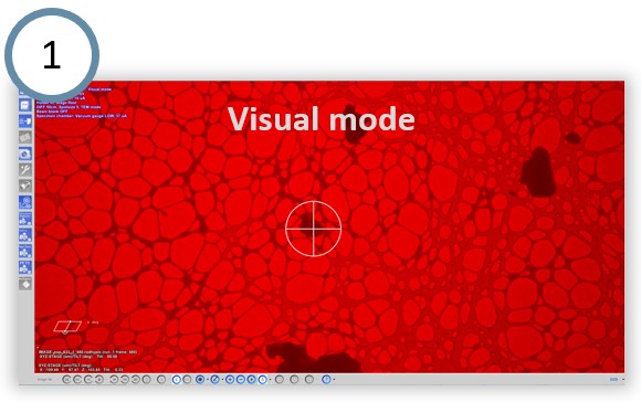
Search for crystals in the visual mode.
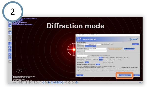
Click the Start experiment button.
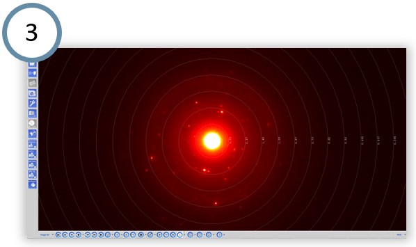
Start the electron diffraction observation automatically.
Structure analysis
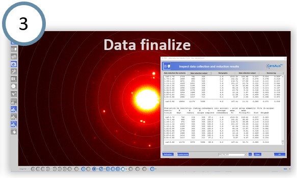
In CrysAlisPro for ED, Autochem performs fully automatic structure determination and refinement during data collection. In addition, it is also possible to manually analyze the structure with efficient and effective functions and merge the dataset obtained from multiple crystals to improve the completeness of the data.
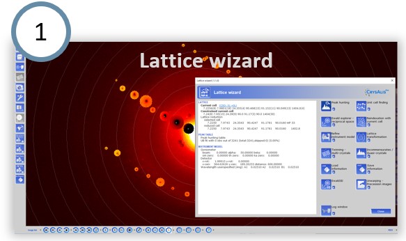
Peak detection and unit cell determination.
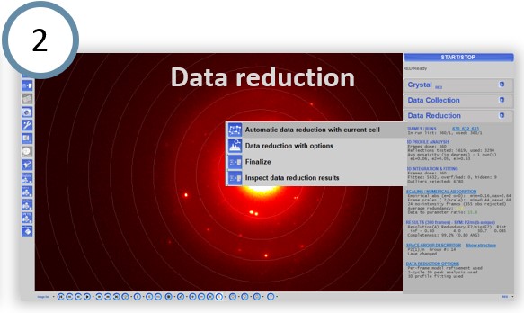
Automatic or interactive data reduction.
Structure refinement
CrysAlisPro for ED allows seamlessly switching to Olex2. The intuitive user interface allows you to build and refine your molecular structure.
Solutions by field
Related products
Related information
Are you a medical professional or personnel engaged in medical care?
No
Please be reminded that these pages are not intended to provide the general public with information about the products.