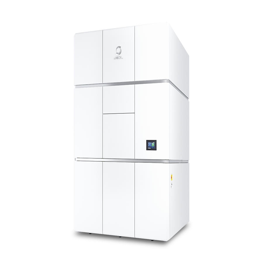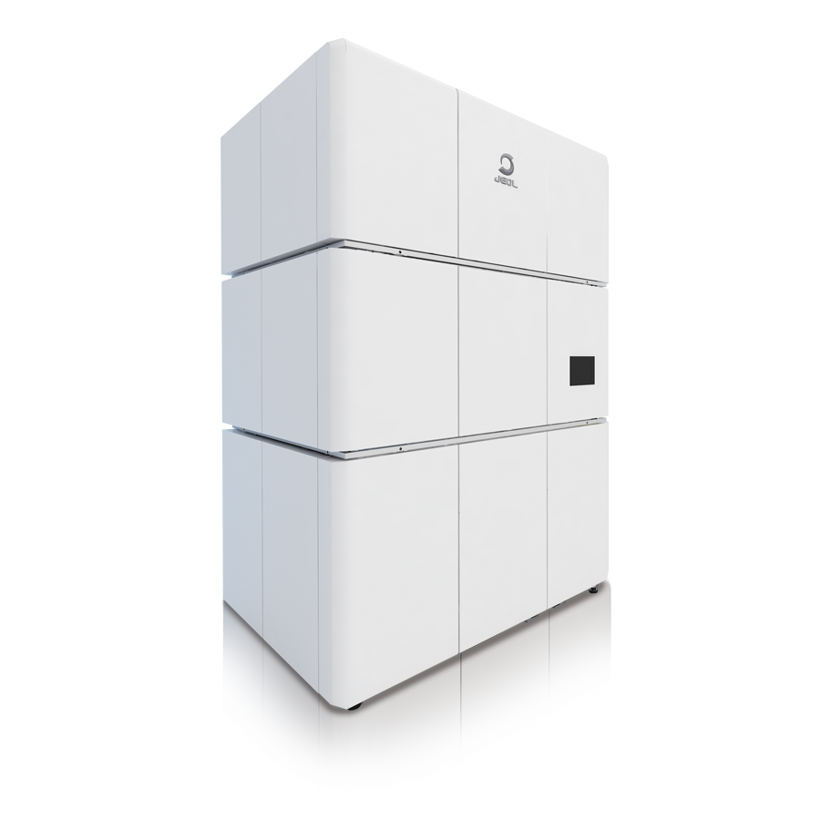


Exploring New Applications of cryo-electron microscopy: Expanding into Soft Materials
INTERVIEW 17
Ayaka Harada, Assistant Professor
TARA, University of Tsukuba
Since the 2017 Nobel Prize in Chemistry recognized the development of cryo-electron microscopy (cryo-EM) for
high-resolution structural analysis, the technique has gained widespread adoption and become an
indispensable tool for studying biomolecular structures. In 2022, two cryo-electron microscopes, the JEOL
CRYO ARM™ 300 II and CRYO ARM™ 200, were installed at the Life Science Center for Survival
Dynamics, Tsukuba Advanced Research Alliance (TARA), as part of an AMED project. A research team led by
Professor Kenji Iwasaki has since achieved significant results in the structural analysis of biological
samples, including proteins and viruses.
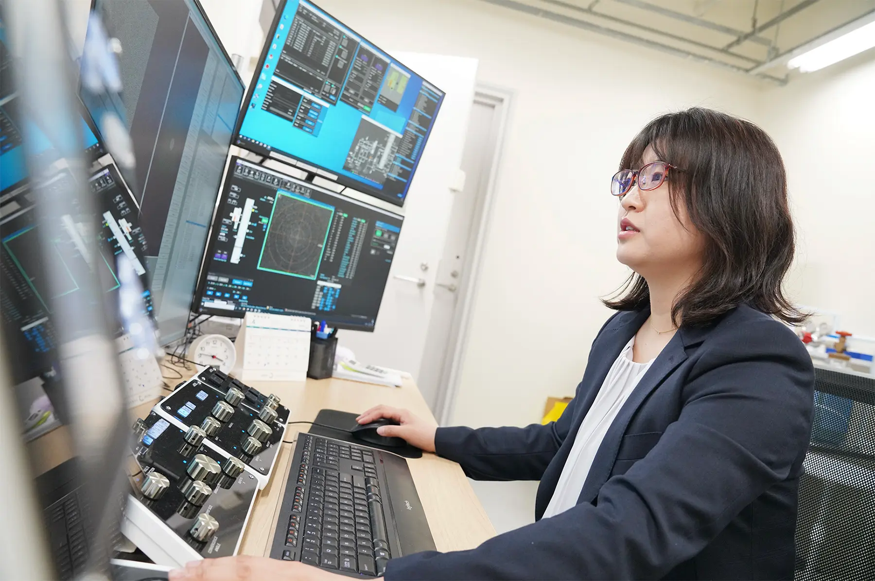
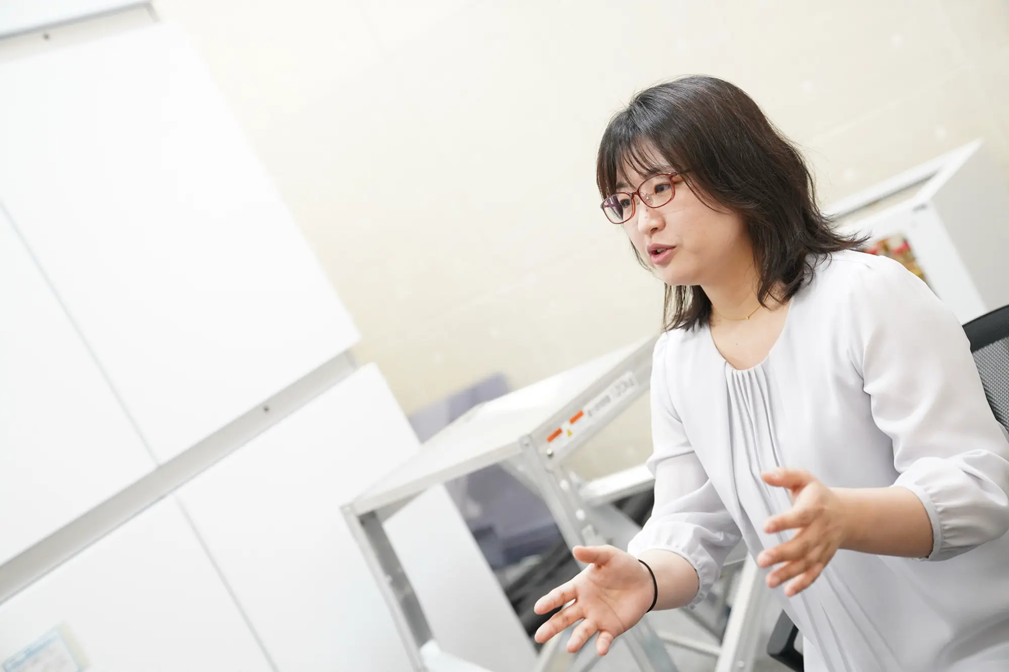
As a member of the team, Assistant Professor Ayaka Harada oversees the shared use of the CRYO ARM™ 300
II. She has been exploring the potential of cryo-EM to observe "soft materials," which have not
traditionally been a primary target of cryo-EM, and is working to expand the technique into new areas of
application.
Cryo-electron microscopy Expands into New Fields as Its Advantages Become More Widely Recognized
Ayaka Harada, who was appointed assistant professor at TARA in September 2021, was assigned to oversee the
shared use of the CRYO ARM™ 300 II, building on her experience in structural biology using X-ray
crystallography.
At first, most measurement requests from companies and external universities involved single-particle
analysis of biological samples, such as proteins related to drug discovery. As the results of these
structural analyses became more widely known, an increasing number of researchers came to recognize the
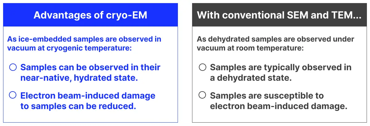
advantages of cryo-electron microscopy, which enables hydrated samples to be observed under vacuum at
cryogenic temperatures. This contrasts with conventional SEM and TEM, in which samples are typically
observed under vacuum at room temperature after dehydration.
As a result, companies that had previously been limited to observing dried samples by SEM or TEM began
approaching us with questions such as, "Can cryo-EM capture clear images of samples in a state closer to
their native condition?"
"When we first started operating the microscope, we mainly focused on single-particle analysis of proteins.
Over time, however, we began receiving inquiries from companies working in materials science about imaging
soft materials, including lipids used in cosmetic products and hair samples. Until then, they had mainly
relied on SEM images, but they were interested in using cryo-EM to obtain new, visually compelling images
with greater impact.
"These were samples I had never worked with before, so I was a little nervous at first. But once the samples
had been sectioned thinly and properly mounted on the grids, we were able to obtain remarkably clear images.
That was when I realized that cryo-EM could also be a powerful tool for visualizing the structures of soft
materials."
Successful Imaging of the Fine Internal Structure of Soft Materials
cryo-EM images revealing the fine structures of proteins are now widely available. But what can we see when soft materials are examined using a high-end cryo-electron microscope? Let's take a look at some actual images.
Cryo-EM images of lipid nanoparticles
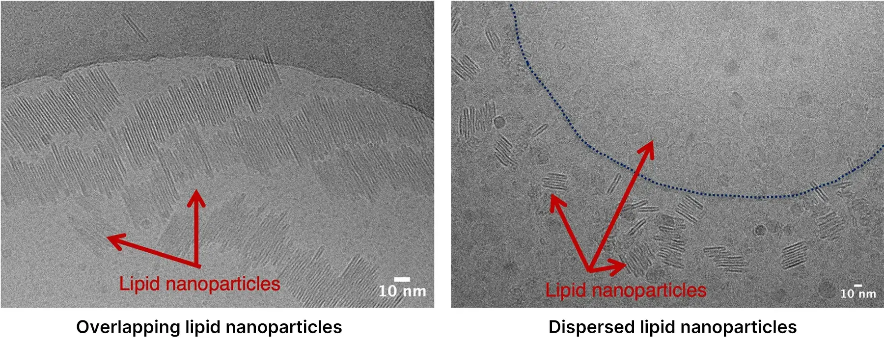
*Image courtesy of Nippon Menard Cosmetic Co., Ltd.
These cryo-EM images show lipid nanoparticles used in cosmetic materials provided by a company and imaged using the CRYO ARM™ 300 II. By minimizing deformation caused by drying or staining, we were able to visualize the native morphology and internal structure of the lipid nanoparticles in a near-native, hydrated state.
Cryo-EM Images of the Microstructure of Damaged Hair
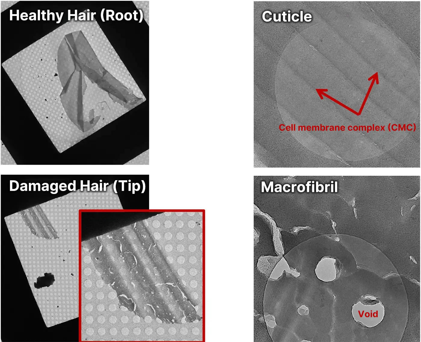
*Image courtesy of NISSAN ARC, Ltd.
Hair, which naturally contains moisture within its structure, is another type of sample that can benefit
greatly from cryo-EM imaging. The images above were obtained in response to a request from a company that
had previously observed dried hair samples by TEM and wanted to examine their structure while preserving
their moisture.
In these images, the structures show no obvious deformation associated with conventional drying or fixation
procedures. Fine internal features can be clearly visualized, including the cell membrane complexes in the
cuticle and numerous voids within the macrofibrils.
Observation image of lipstick (lipid sample)
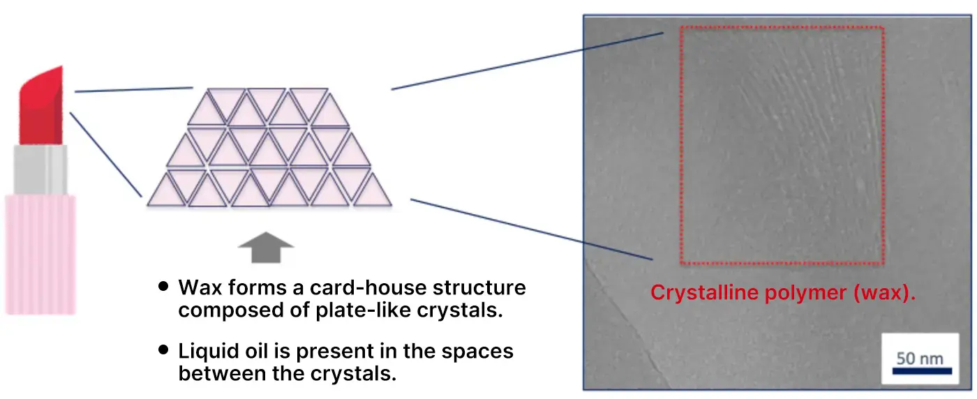
*Image courtesy of NISSAN ARC, Ltd.
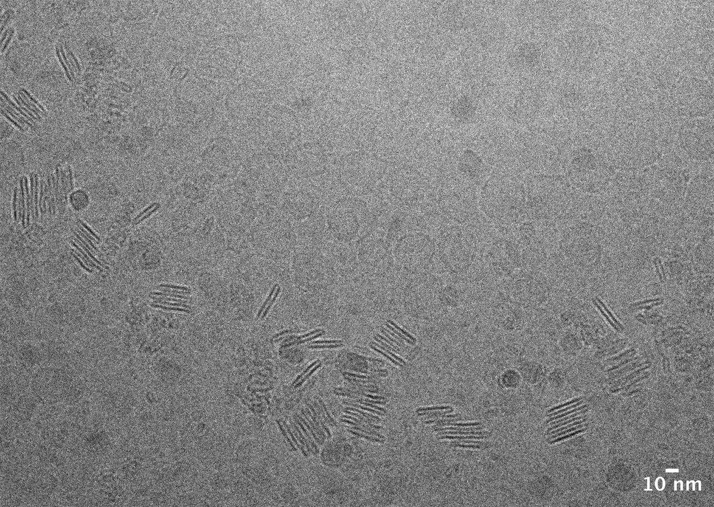
This cryo-EM image shows the microstructure of lipstick. In lipstick, wax crystals form a card-house
structure, with oils and pigments filling the spaces between the crystals. The smoothness experienced during
application is related to how this structure breaks down as the lipstick is applied. cryo-EM was therefore
used to examine the fine structure of the card-house arrangement and how the crystals are stacked.
The image shows regularly stacked crystals without obvious structural disruption from electron beam damage.
This demonstrates that even samples containing oils and other soft components can be examined by cryo-EM
while minimizing deformation, allowing their internal structures and the distribution of different
components to be visualized.
As the usefulness of cryo-EM for investigating the fine structures of soft materials became increasingly
apparent, the number of requests for cryo-EM analysis began to rise rapidly.
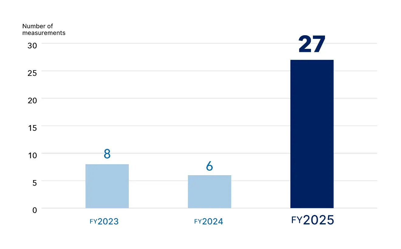
Trend in the number of soft materials measurements
Cryo-EM Reveals Emerging Needs in Industry
Through discussions with companies involved in these projects, we gradually began to gain a clearer picture
of what industry is looking for. There continues to be strong demand for single-particle analysis of
proteins and viruses. At the same time, as cryo-EM has become more widely accessible, a previously hidden
need has begun to emerge: the desire to visualize the fine internal structures of soft materials in a state
as close as possible to their native condition. In other words, there were many people looking for ways to
see structures that conventional SEM and TEM could not reveal.
Similar trends appear to be emerging beyond TARA. For example, we have received a number of inquiries from
the National Institute for Materials Science (NIMS), also located in the Tsukuba area, about observing
aqueous materials by cryo-TEM rather than SEM. We have also heard of facilities where cryo-electron
microscopes are so heavily booked for biological applications that materials-related projects have had to be
put on hold.
"We've been receiving inquiries from some rather unexpected fields as well. One of the latest examples for
me was a request to see whether we could observe a suspension of clay-like minerals. I remember thinking,
'Geoscience now, too!' I was surprised, but since it is an aqueous sample, it looks like we may be able to
apply the same kinds of sample-preparation and imaging approaches that we have traditionally used for
single-particle cryo-EM.
"I find that quite exciting. Unexpected requests like this may lead us to discover entirely new ways of
using cryo-EM to approach scientific questions."
Taking on the Challenge of Automated Morphological Quantification through Image Analysis
At this early stage of soft-material imaging by cryo-EM, a measurement may be considered successful if even
a single striking image clearly reveals the fine internal structure of the sample, or if the images provide
a qualitative indication that a particular target morphology is abundant.
However, Assistant Professor Harada believes that the analysis of soft materials needs to become more
quantitative. For example, do the morphologies frequently observed on a particular grid truly represent the
sample as a whole? What proportion of the observed area do they occupy? Addressing questions like these will
be important for improving the reliability of soft-material analysis.
Particles with diverse morphologies observed for lipid samples
*Image courtesy of Nippon Menard Cosmetic Co., Ltd.
"When we obtain an image showing an interesting morphology, I would like to automatically determine what
proportion of the entire grid image is occupied by that morphology. In single-particle analysis, we
routinely collect thousands of images, and I think we could apply that experience to soft materials. By
collecting large numbers of images, we could evaluate the distribution of different structures and
automatically quantify specific morphologies.
"The soft-material samples brought to us are often heterogeneous and contain a variety of components and
structures. If we could classify and quantify them based on their morphology, I believe that would be very
useful for interpreting the results.
"Liposomes, for example, are often used to encapsulate drugs. With cryo-EM, we can distinguish between
liposomes containing a drug and empty liposomes based on their morphology. If we could collect large numbers
of images and automatically analyze them to determine drug encapsulation efficiency, I think that would be
extremely useful. Developing this kind of automated image analysis is something I would really like to take
on."
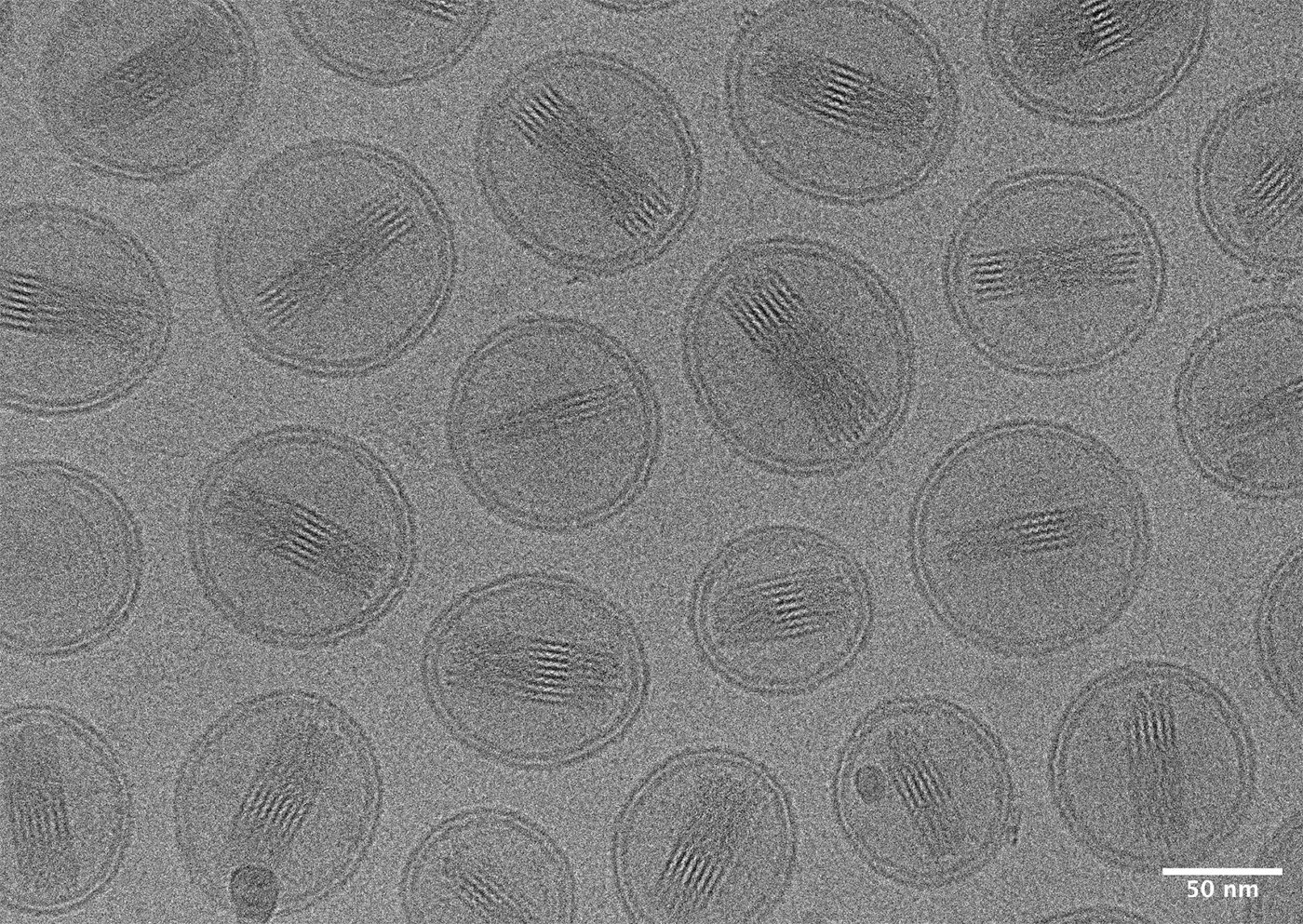
Liposome with a drug inside
Collaboration Beyond the University—and with SEM and TEM Researchers—Is Key
We asked Dr. Harada, who is exploring new applications of cryo-EM for soft materials, what she believes will
be the key to making these efforts successful.
"I think collaboration beyond the university will be absolutely essential.
"For example, I believe there is a great deal to be gained from collaborating with the National Institute
for Materials Science (NIMS), which has a wide range of electron microscopes and works with an enormous
variety of materials. By bringing together researchers with expertise in SEM and TEM and our expertise in
cryo-EM, we can examine the diverse materials available at NIMS and continue building our knowledge of where
cryo-EM offers unique advantages. In doing so, I believe we can create an environment in which cryo-EM is
more accessible not only to researchers in biology, but also to those in materials science.
"I would also like to expand these collaborations to include research institutes such as AIST and KEK. By
working together across the Tsukuba area and establishing the region as a hub for soft-material imaging, I
hope we can develop a distinctive strength that sets us apart from other cryo-EM facilities."
The Challenge of Soft-Material Imaging: Sample Preparation
Recently, we have seen a rapid increase in requests to examine ionomers in fuel-cell catalyst layers.
Because these ionomers are dispersed in solution, they can be observed by cryo-EM in much the same way as
proteins in single-particle analysis, by embedding them in a thin layer of vitrified ice.
Solid samples, on the other hand, present sample-preparation challenges that are not encountered in
conventional single-particle analysis. Samples such as the hair, lipstick, and other soft or semi-solid
materials shown above require different preparation methods and may need to be frozen and sectioned into
thin slices. The sections must then be mounted on grids, transported to the cryo-electron microscope while
maintained at cryogenic temperature, and loaded into the microscope cartridge. If the temperature rises
during this process, the sample may be damaged, making imaging impossible. Once the sample has been
successfully prepared and loaded, however, the subsequent imaging and analysis procedures are not
substantially different from those used for protein samples.
In other words, the biggest challenge at present lies in sample preparation: how to section a frozen solid
sample efficiently and transfer it into the cryo-electron microscope while maintaining cryogenic conditions
throughout the process.
"The solid samples we are currently observing are sectioned at other facilities and then brought to us.
Until we improve our own sample-preparation capabilities in this area, it is difficult to confidently say,
'We can image soft materials at TARA!'
"Ideally, we would have a system that allows frozen samples to be thinned using an ion beam--for example,
with a JEOL cryo-FIB--and then transferred directly to our JEOL CRYO ARM™ 300 II. That would reduce the
risk of failure considerably. Cryo-FIB systems, however, are extremely expensive and not something we can
easily introduce on our own.
"This is another reason why I believe collaboration with organizations in the Tsukuba area, including NIMS,
is so important. It may be difficult for the University of Tsukuba alone to introduce such expensive
equipment, but if we approach it as an 'All-Tsukuba' initiative, there may be possibilities.
"That said, I can't simply wait for someone else to make it happen, so I've been working on a few ideas of
my own!"
Tackling the Challenge through an "All-Tsukuba" Initiative
One might expect a researcher simply to hope that "perhaps one of the nearby research institutes will
install a cryo-FIB someday." But Harada prefers to take action herself and work toward making that
possibility a reality.
In April, she applied for a one-year pilot project in collaboration with soft-material researchers at the
National Institute for Materials Science (NIMS), tentatively titled "Microstructural Analysis of Materials
Using Cryo-Electron Microscopy."
"If the project is accepted, I would first like to examine a wide range of samples for which high-quality
TEM images have already been obtained, and systematically compare what we can see by TEM and cryo-EM. Based
on the knowledge gained from these observations, I would then like to pursue larger competitive research
grants and expand the project further.
"If, along the way, we could introduce sample-preparation equipment such as a cryo-FIB somewhere in the
Tsukuba area, we might eventually be able to establish Tsukuba as a place people think of when they want to
study soft materials. That's one of my ambitions!" she says with a laugh.
"Personally, I would like to establish a high-throughput cryo-EM platform for efficient structural analysis
of soft materials. My goal is to develop an integrated workflow that brings together sample preparation,
imaging, and image analysis, and ultimately establish it as a versatile structural-analysis platform that
can be routinely used by researchers studying soft materials and other materials whose structures function
in solution or under hydrated conditions."
We look forward to seeing what comes next from Assistant Professor Ayaka Harada, a researcher who brings together expertise in diverse technologies—including SEM, FIB-based sample preparation, and cryo-EM—through cross-disciplinary collaboration, while engaging researchers across different communities to explore new scientific approaches and build a distinctive identity for the TARA Center.
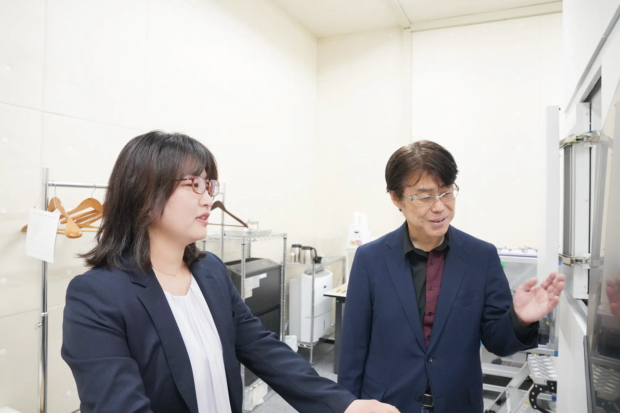
<Iwasaki Laboratory, TARA, University of Tsukuba>
The laboratory is led by Professor Kenji Iwasaki, who heads the Structural Life Science Research Project at TARA. Using structural biochemistry approaches, the laboratory conducts research on topics including the structural analysis of molecules involved in soft tissue tumors, as well as the operation of state-of-the-art cryo-electron microscopes, including the CRYO ARM™ 300 II and CRYO ARM™ 200 (JEOL Ltd.).
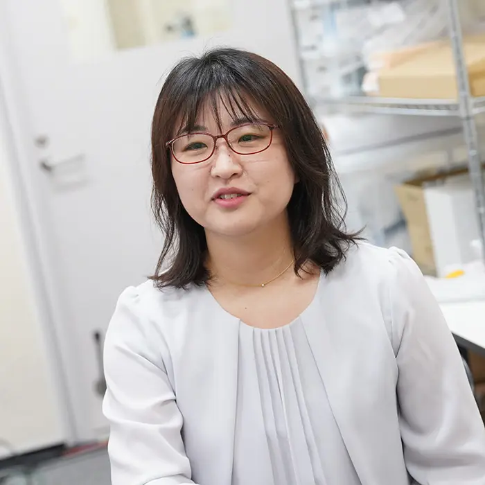
Ayaka Harada
Iwasaki Laboratory, TARA
She received her Ph.D. from the Graduate University for Advanced Studies (SOKENDAI). After working as a researcher at the Institute of Materials Structure Science, High Energy Accelerator Research Organization (KEK), and as an Assistant Professor at the Faculty of Pharmacy, Keio University, she joined TARA at the University of Tsukuba in September 2021 and has held her current position since then.