

Making MicroED, the Ultimate Tool for Crystal Structure Determination, Easier to Use!
INTERVIEW 16
Naruhiko Adachi
Associate Professor
Life Science Center for Survival Dynamics,
Tsukuba Advanced Research Alliance (TARA),
University of Tsukuba
A method for elucidating the structures of crystals smaller than 1 µm - too small to be analyzed even with synchrotron X-rays - was proposed by several researchers in the 2013-2015 timeframe, using electron diffraction data. This is the MicroED (Micro Electron Diffraction) method using cryo-EM (cryo-electron microscope), a breakthrough technique that enables observation of the chemical structures in specimens where trace and polymorphic crystals are present for the first time. Associate Professor Naruhiko Adachi of the Life Science Center for Survival Dynamics at the Tsukuba Advanced Research Alliance (TARA) has been promoting the shared use of cryo-EM as dedicated instrument for MicroED, while working to establish electron diffraction workflows.
A MicroED-Dedicated Instrument Established at TARA!
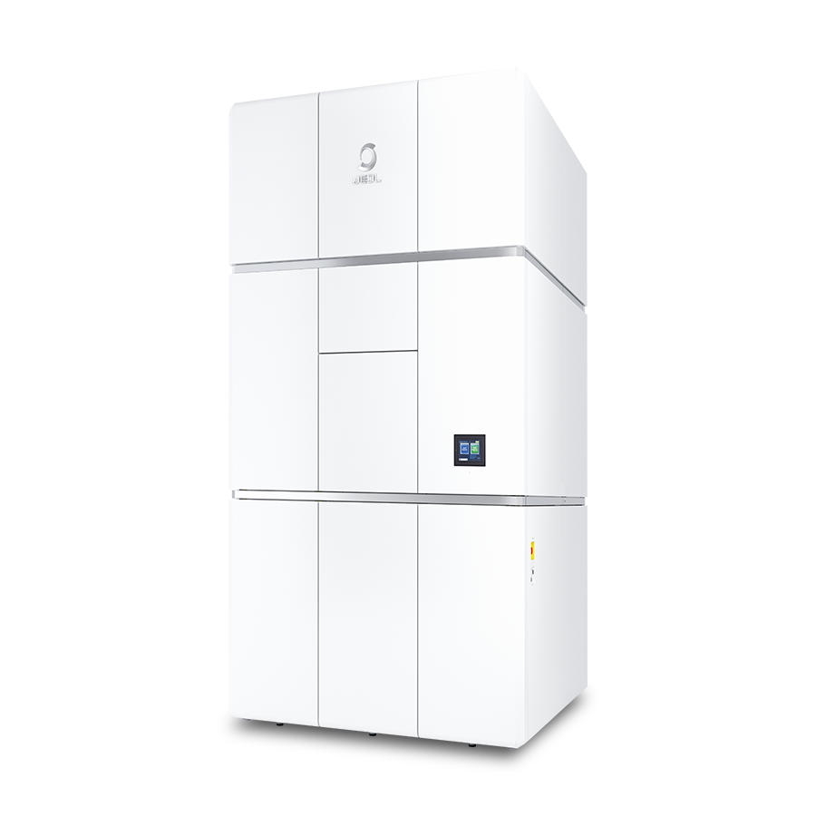
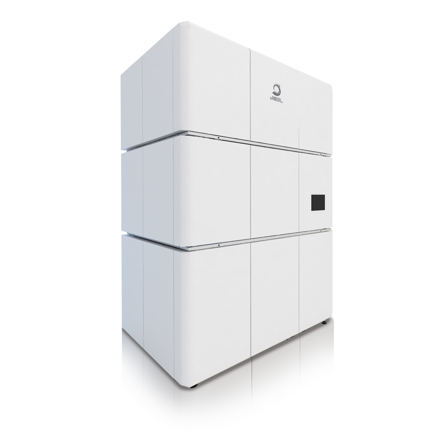
In 2018, a cryo-EM was introduced to High Energy Accelerator Research Organization (KEK), Inter-University Research Institute Corporation, which specially appointed assistant professor (then) Naruhiko Adachi, who had been conducting research about gene expression regulation, belonged to. Since he had experience using a cryo-EM in another facility, he was entrusted with managing the instrument. He made efforts to expand the use of single-particle analysis by preparing manuals for the workflow. From 2019, he also began working on MicroED and tried to introduce MicroED experiment with the cooperation of an expert in X-ray crystal structure analysis in KEK, Yusuke Yamada, then Assistant Professor. Following that, in 2023, Dr. Adachi took the post of an associate professor at TARA where two cryo-EMs (CRYO ARM™ 300 II and CRYO ARM™ 200 made by JEOL Ltd.) had been installed. Since then, he has introduced MicroED experiments and steadily accumulated measurement experiments.
"When I joined TARA in 2023, Dr. Iwasaki and Dr. Harada at TARA were already operating the CRYO ARM™ 300 II for single-particle analysis. The CRYO ARM™ 200 which had been installed at the same time was made available for us to use as a dedicated MicroED instrument."
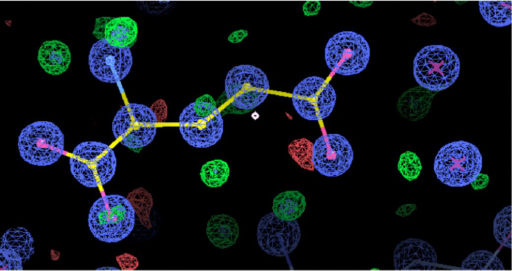
Crystal structure of a molecule (sodium glutamate) obtained by MicroED measurement.
Smooth MicroED Measurement with a Dedicated Instrument
MicroED at TARA has two distinctive features:
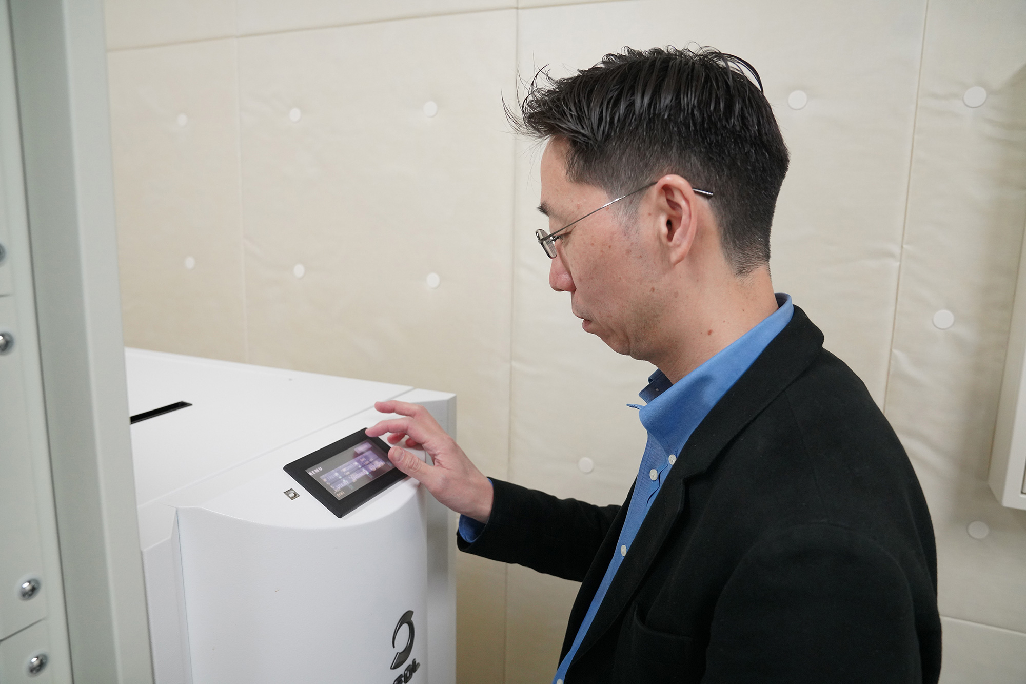
One is that the CRYO ARM 200 is operated as a dedicated instrument for MicroED. While it is possible to use a single cryo-EM for both single-particle analysis and MicroED measurements, the measurement conditions must be reconfigured each time the system is switched between the two methods. Because single-particle analysis and MicroED require different electron dose conditions, switching between them takes a considerable amount of time. At TARA, however, the CRYO ARM 200 has been operated as a dedicated MicroED instrument since its installation, enabling smooth and efficient operation. A cryo-EM shared and operated exclusively for MicroED is rare even on a global scale. Its existence is highly valuable and represents one of TARA's major strengths.
"From the perspective of facility operation, cryo-EM instruments are often managed primarily for single-particle analysis, which remains the dominant measurement technique. As a result, there are very few cryo-EMs dedicated exclusively to MicroED. Therefore, being able to operate the CRYO ARM 200 as a dedicated MicroED instrument is extremely valuable, in terms of both convenience and operational stability."
The Key to Ease of Use - an Automated Workflow
The second distinctive feature is its "ease of use".
There is certainly a demand for MicroED, which enables extremely small, trace, and polymorphic crystal structures. However, it is not as easy as single-particle analysis where the workflow and software are already well established. Nevertheless, Associate Professor Adachi at TARA handles MicroED measurement requests for around 40 research groups annually. What is the mechanism behind that?
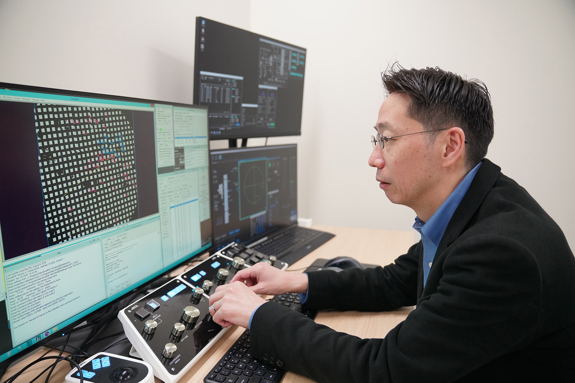
"Because the cryo-EM we operate is a shared facility, it needs to be easy to use for outside users. Since most outside users only access it once every one or two months, the use needs to be as user-friendly as possible. For this purpose, we believed that it was necessary to establish a workflow as automated as possible and to prepare a comprehensive manual. However, there are limits to what members of a single facility can accomplish alone. So, a MicroED on-line community was launched for more than one facility to collaborate. We introduced automation software with the help of Dr. Makino of Osaka University, Dr. Yanagisawa of the University of Tokyo, and Dr. Nakane of Osaka University. The processing of measured data was prepared by Dr. Yamada of Tohoku University with the help of Dr. Nakane of Osaka University and others. Once the analytical system for automated measurement and data processing was ready, we repeatedly used it with the dedicated instrument at TARA and
increased the degree of completion, while we also worked on preparing the manuals.
In this way, we were able to prepare the automation of measurements and data processing as well as the manuals, the MicroED at TARA became easy to use even for the outside users. "
Is MicroED at TARA Easy Enough for Even Senior Undergraduate Students!?
According to the efforts, even students can measure about 200 particles for a night, 1000 particles over the weekend, if they follow TARA's MicroED workflow which has been automated as much as possible. In experiments, which require the determination of guanine crystals of fish, students in their senior undergraduate program even accomplished the measurements of about 2000 particles in a week. It is rare case worldwide for senior undergraduates to fully use MicroED instruments with the cryo-EM instrument. This is because the workflow incorporates automated measurement and data processing, enabling stable and reliable operation.
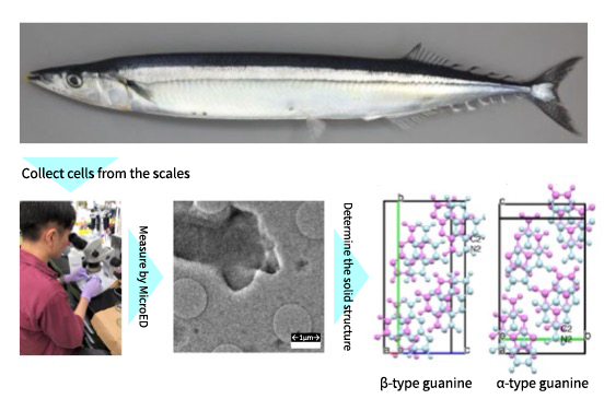
Senior undergraduate students measured fish (Pacific saury) and confirmed that β type and α type guanines were contained!
The measurement results for the guanine crystals along with the results of MicroED-workflow automation, are being submitted as an original paper.
"It has been known that fish scales are shiny in silver or blue, because there are guanine crystals in the fish skin. But whether the guanine is α-type or β-type, was not known. The guanine crystal in the scale was too small to determine the crystal structure using X-ray diffraction. In 2024, it was discovered that the guanine structure of salmon was β type. That led us to question if other fish species also have β-type guanines, or α-type, or possibly, polymorphic crystals consisting of β-type and α-type. Therefore, we decided to examine the cell structures at the root of the scale for Pacific saury, cutlassfish, and sapphire damselfish with senior undergraduate students. MicroED can precisely determine the crystal structure, even if the specimen has β-type and α-type, a polymorphic crystal is mixed, as it observes individual crystals in the specimen. However, in case a crystal is taken from a cell, the crystal can be
damaged or mixed with fish flesh. We can hardly find the target crystal unless we perform numerous measurements of uncertain particles. As a result of measuring approximately 2000 particles, we could confirm the existence of guanine crystals of both β-type and α-type. I think this is thanks to the enthusiasm of excellent students as well as advanced automation of measurement and data processing.
As a side note, I also had the students obtain the fishes for experments. As the fish lose freshness, the crystals also deteriorate. So, I am sure that the students must have developed the ability to spot "fresh, lively Pacific saury. (laughs)"
MicroED Fills the Gaps in the Coverage of Other Analytical Methods
MicroED can obtain diffraction data even from a crystal less than 1 µm in size in a specimen consisting of trace and polymorphic crystals. However, it is difficult to measure a specimen containing a mixture of various particles when it is unclear whether the target crystal is present. Using MicroED alone, it is difficult to determine how many particles need to be observed, whether 100 or as many as 1,000. In such cases, it is effective to check if the target crystal is present in the specimen by using powder X-ray diffraction, then to analyze the structure with the MicroED.
Moreover, the data measured with the MicroED reveals the structure (form) of the molecule but does not identify the kinds of elements. MicroED alone cannot analyze the structure of completely unknown substances. If the structural formula and elemental composition are known, each atom can be identified based on that. Therefore, combination with mass spectrometry and NMR is useful. Thus, to take advantage of MicroED for molecular structure determination, it is important to combine it with other analytical methods.
"One research group had been trying for a long time to determine the structures of compounds that existed only as tiny crystals and in extremely small quantities, using powder X-ray crystallography. However, even after two years, they could not identify any of the structures. Despite spending all that time improving the crystal quality, powder X-ray crystallography alone could not solve any of the structures. At their wits' end, they tried MicroED at TARA, then they managed to determine the structures of 10 crystals in just one week! The whole research group was celebrating like a festival (laughs). We have had several experiences like that. By measuring the improved crystal specimens with MicroED, the structures were solved all at once. This is a great example of what the combination of powder X-ray crystallography and MicroED can achieve. Even if the crystallinity is poor at first, as long as powder X-ray crystallography indicates that the target crystal is present, the crystal quality can be gradually improved. Then, each individual microcrystal can be analyzed one by one using MicroED. I think this kind of workflow is extremely well suited to MicroED."
While MicroED has groundbreaking strengths, it also has limitations when it comes to analyzing completely unknown substances. In this sense, this is a technology that fills the gaps in the coverage of existing three-dimensional structure analysis methods. In order to maximize its capabilities, complementary use with powder X-ray diffraction, mass spectrometry, and NMR are highly effective.
The Key to Success of MicroED is In-Advance Meeting and Attendance during the Measurement
When MicroED is used at TARA, an in-advance meeting is the most important to succeed in the measurement. Ideally, the measurements should be performed based on a clear shared understanding of factors such as the specimen's conditions and characteristics, the data analyst's expertise, the purpose of the analysis, the requestor's knowledge of crystal analysis, and the technical limitations of MicroED.
"If we start the measurement after thorough information sharing with the requester, we often have a smoother measurement. Of course, after examining what the requestor wants to know, we sometimes decline the measurement request as it is not possible with the MicroED. In addition, it is extremely important that someone who is familiar with the particular specimen attend during the measurement. It is troublesome if only the specimen is provided. We do not know much about the specimen. If something happens during the measurement, we need someone who knows the specimen well to make judgements for various matters, in order to make the measurement successful. "
We Want to Establish a National Center for MicroED!
Since 2023, Associate Professor Adachi has been working on the theme of "Standardization of MicroED through Automation", in collaboration with many researchers. We asked him, the expert of MicroED who knows its advantages and disadvantages about his future ambitions.
"MicroED is still a new technology and there are many areas where we are feeling our ways. In addition to ensuring the smooth operation of cryo-EM at TARA as a shared facility, we would like to continue refining the operation system by listening carefully to user's feedback in real-time and utilizing the advantages of having a dedicated instrument. Then, just as we have already exported the TARA system to other research institutions such as Osaka University and SPring-8, we hope to actively expand the system to outside research institutions. Ultimately, we would like to establish a "National Center for MicroED" with about 10 cryo-EMs of the same level as TARA, so that we can support even more researchers in the future!"
Naruhiko Adachi
TARA in University of Tsukuba (Dual Appointment: JEOL Ltd.)
After serving as a Special Researcher at the Japan Biological Informatics Consortium and as a Specially Appointed Associate Professor at the Institute of Materials Structure Science, High Energy Accelerator Research Organization (KEK), he has held the current position since December 2023. Ph.D. (Pharmaceutical Sciences).