Ultra-fast analysis of protein 3D structures
― preparation of a sample for
cryo-EM in 10 minutes ―
INTERVIEW 15
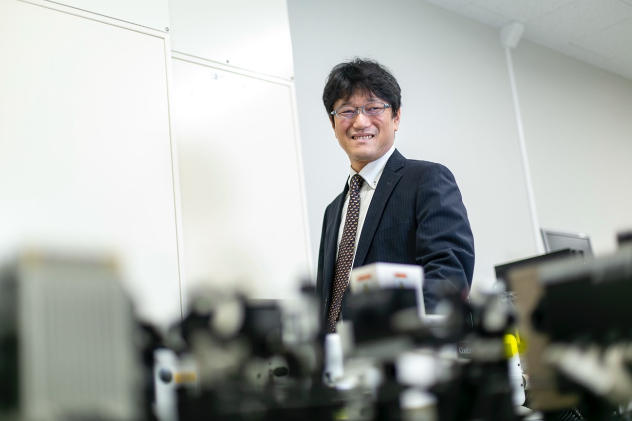
Tsuyoshi Inoue
Laboratory of Structure and Function Analysis of Biomolecules
Graduate School of Pharmaceutical Sciences, The University of Osaka
The 3D structure of a biomolecule, such as a protein is becoming an essential information for research and development of biochemistry and drug discovery. While the analysis method using cryo-EM is widespread, a tool to dramatically improve the speed of analysis work, "EG-grid™" was developed by Prof. Inoue. We interviewed Prof. Inoue about its characteristics.
Rapid Penetration of cryo-EM
As a method for 3D structural analysis of proteins, cryo-electron microscopy (cryo-EM) has rapidly spread. In the international public database PDB (Protein Data Bank), 3D structure data of biomolecules (proteins, nucleic acids, etc.) that are newly elucidated are registered day by day. The data is disclosed once the related paper is released. Among the structural data disclosed, the ones elucidated by electron microscopes reached 5,791 in a year in 2024. This occupies 37% of the total number of disclosed data in that year.
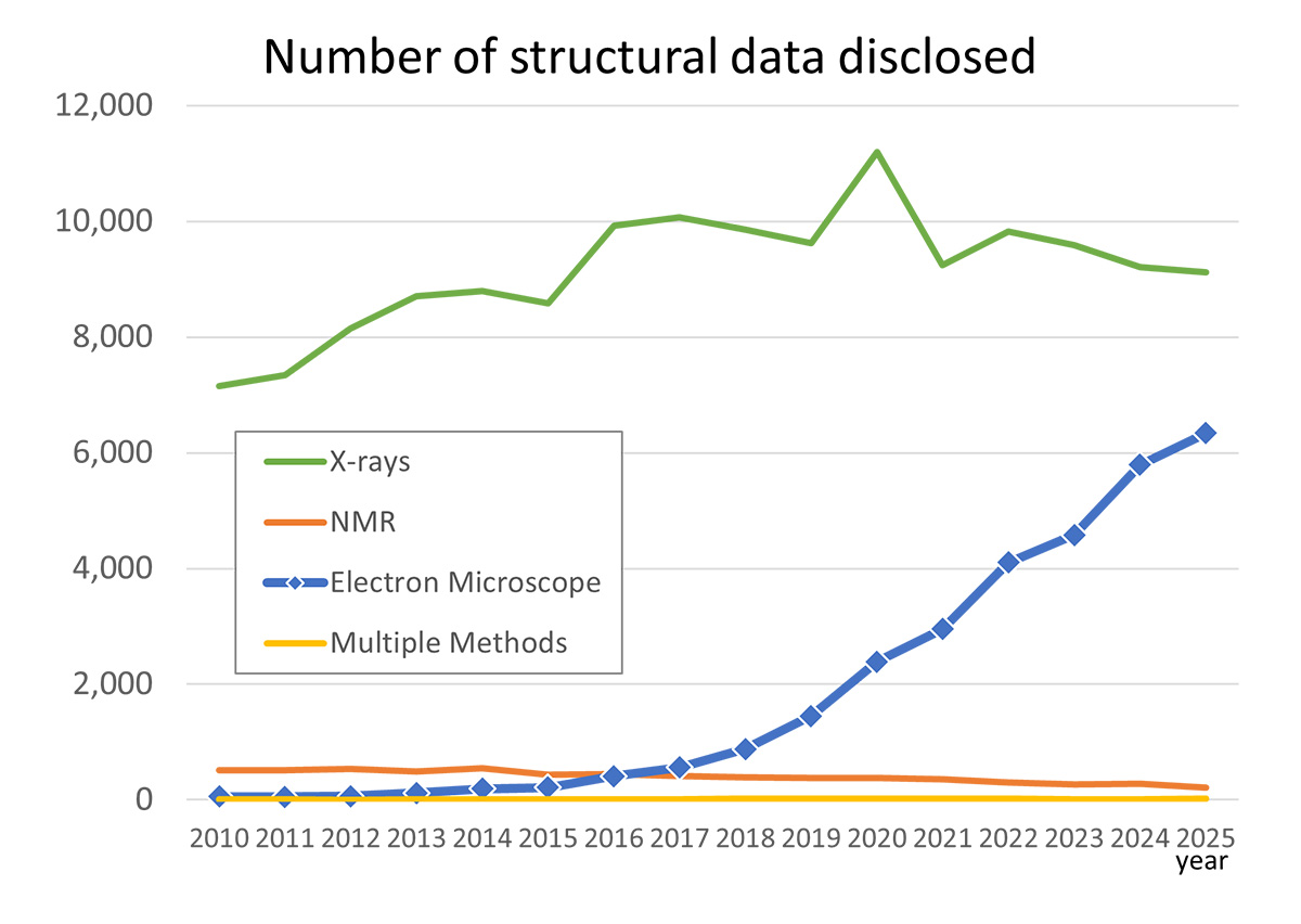
Number of disclosed PDB by analysis method in a year. Created based on statistical data available with PDB. The record in 2025 is as of December 3.
According to the graph, the data using electron microscope began increasing around 2017.
This year, the three researchers who developed the fundamental technology of the cryo-electron microscope
were awarded the Nobel Prize in Chemistry. "Triggered by the Nobel prize, the awareness of cryo-EM and its
method was increased, accelerating the use", says Prof. Tsuyoshi Inoue (Graduate School of Pharmaceutical
Science, The University of Osaka).
The reason for the popularity of the cryo-EM is that the crystallization of protein, which used to be an
inevitable process in an X-ray crystal structure analysis, is not required. The X-ray crystal structure
analysis is a major method for elucidating the 3D structure. However, in this method, protein needs to be
made into a large crystal of about 100 μm. Sample preparation takes several months or more. Also, there are
proteins and complexes that are hard to crystalize. The cryo-EM can analyze proteins that are hard to
crystalize, which is the advantage of the cryo-EM.
Moreover, analysis is possible even if there are only small sample amount available, which is another great
advantage. Challenging structure analysis becomes easy even for proteins whose purification is difficult and
a complex which is unstable and tends to easily aggregate.
Sample preparation in a month even for the cryo-EM
Then, does cryo-EM require any processing? Yes, it does. In the early stage, sample preparation and taking electron micrographs took some time.
For analysis of a 3D structure, a single-particle analysis method is used. In this method, many images of a discrete single particle, are taken by the cryo-EM. To increase accuracy, many images taken from various angles need to be prepared and they are reconstructed into a 3D structure using a computer. For this purpose, it is necessary to prepare an appropriate sample which allows efficient image acquisition, thereby improving the overall research speed.
For preparing a sample, the ice-embedding method was traditionally used. This is a method to rapidly freeze a solution to embed proteins within amorphous ice. However, this method presented challenges in adjusting the concentration. If the concentration is too high, particles overlap and usable images cannot be obtained. On the other hand, if the concentration is too low, the number of particles decreases, and imaging efficiency is reduced. In addition, if the ice is thick and protein particles are distributed in the thickness direction, the number of particles that can be focused is limited, further reducing imaging efficiency.
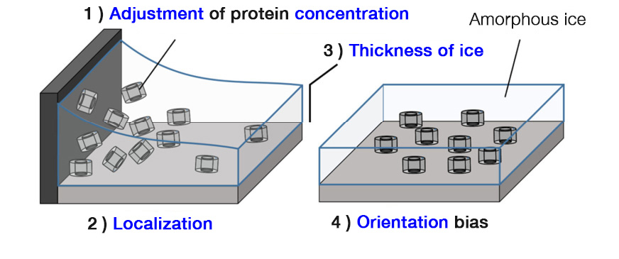
Issues in sample preparation by ice-embedding method
"Adjusting concentration is a tough thing. However, the most critical problem in the ice embedding method is the preferred orientation, where the proteins align in the same direction!" (Prof. Inoue)
The preferred orientation is generated during freezing processes. But in some cases, the orientation to the same direction occurs before freezing. These are the cases where highly hydrophobic proteins adhere to the gas-liquid interface, resulting in orientation in the same direction. In the conventional sample preparation method, it could take a month to consider these conditions.
Prof. Inoue and others succeeded in developing a sample preparation tool, "EG-grid™ (Epoxidized Graphen Grid)", a great solution of those issues. EG-grid™ is a support base made of graphene bearing epoxy groups. Since the epoxy group has the property to capture lysine residue existing at the surface of the protein particle, protein particles are adhered at various angles. Epoxy group density is properly pre-set, so there is no need to worry about the concentration adjustment of the solution. There is no distribution in the thickness direction, allowing appropriate focusing on imaging.
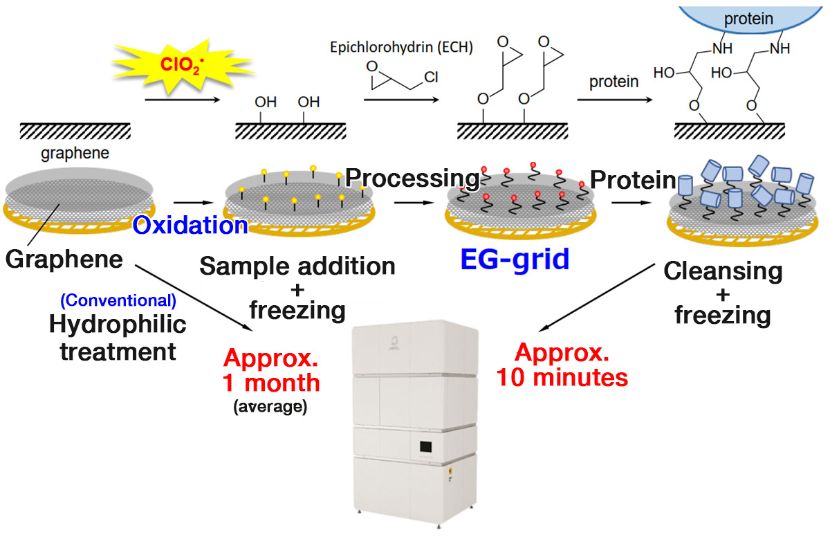
By using the EG-grid™, sample preparation time which previously took nearly a month has been reduced to about 10-15 minutes. A sample can be set on a cryo-electron microscope within 20 minutes even including the freezing process.
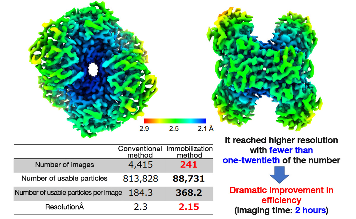
The imaging efficiency has also improved. The number of usable particles obtained from a single photographing has increased compared with the ice-embedded method, reducing the total number of images required. For some proteins, analysis can even be completed with as few as one-tenth or one-twentieth of the conventional number of images.
Structural analysis example of Protein-B
Comparison of imaging efficiency between conventional method (ice-embedding method) and the immobilization
method (using EG-grid™)
Development triggered by "mysterious disinfectant/deodorant"
The development of EG-grid™ began with an encounter with a "mysterious disinfectant/deodorant".
In 2015, an inquiry was raised to the University of Osaka. "When a terminal oral cancer patient used this
disinfectant/deodorant to control odor, the cancer appeared to regress. Could you investigate the underlying
mechanism? If the effect is real, we would like to turn it into an anticancer drug." Along with this
request, a bottle of the "mysterious water" was handed over.
We asked, "What are the ingredients?" The answer was only, "We cannot disclose that."
It sounded rather dubious. However, when the analysis was entrusted to Prof. Shunichi Fukuzumi of the
Graduate School of Engineering (at that time), a specialist in radicals quickly identified the principal
component and the underlying mechanism.
The principal component was the chlorite ion (ClO2-). It turned out that this ion (ClO2-) produces reactive chlorine dioxide (ClO2) in water as much as required only when there are target bacteria and viruses existing - this was the mechanism.
Later, the University of Osaka decided to call this highly safe "water" MA-T: Matching Transformation System. This is to avoid confusion with other substances that can generate chlorine-based gas.
Specially-appointed Prof. Ohkubo (at that time) who handled the analysis in the same research group as Prof. Fukuzumi, is an expert in photochemistry. He became intrigued: "What will happen if we irradiate with light on this?" Under acidic conditions, he released chlorine dioxide as a gas and exposed it to light. He noticed that it split into reactive oxygen species and chlorine radical. He further found that using these reactive oxygen species and chlorine radical could oxidize various substances.
Professor Dr. Kei Ohkubo
Institute for Open and Transdisciplinary Research Initiatives
The University of Osaka
For example, oxidizing methane generates methanol and formic acid. There is no emission of carbon dioxide. It was the first time in the world to synthesize methanol under the conditions of normal temperature and normal pressure. Methane can be obtained by fermentation of cow manure, or at the collection site of petroleum and natural gas but are difficult to transport. However, by making it into liquid methanol and formic acid, the transportation becomes easy, and its utilization expands.
EG-grid™ is the result of the utilization of such oxidation reaction into the cryo-EM. If the
graphene surface is oxidized to bear hydroxyl groups (-OH), a functional group that can immobilize protein
could be substituted there. This idea was brought into developing the EG-grid™.
Thus, Prof. Inoue thought that newly-found oxidation reaction and highly safe MA-T could be applied to a
wide range of uses, and he applied for OPERA (Program on Open Innovation Platform with Enterprises, Research
Institute and Academia by JST (Japan Science and Technology Agency) for further research on practical
applications. However, for the application to be adopted, a significant amount of joint research funding was
needed. The deadline was less than three months away. One of the companies that responded to his urgent
appeal was JEOL Ltd.
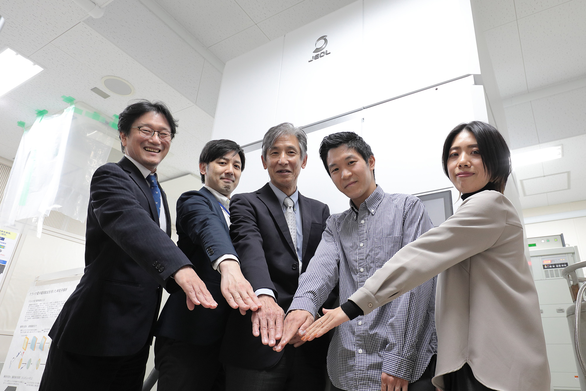
JEOL established "Osaka University-JEOL YOKOGUSHI Research Alliance Laboratories" in 2018, and so the
research funding was provided from these laboratories. The proposal of Prof. Inoue was successfully adopted
for OPERA (for the period of October 2019 to March 2024).
This allows for the development of the EG-grid™ and support of the project by JEOL CRYO ARM™ 200 which was installed in
the laboratories at the same time.
This OPERA project has brought many results and was highly evaluated. In February 2024, it won the Prime Minister's award from the 6th Japan Open Innovation Prize. The research on oxidizing methane received the Commendation for Science and Technology by the Minister of Education, Culture, Sports, Science and Technology in April, 2024. Also, their efforts have been picked up in the proposal by the Science Council of Japan "the collective strength of industry, government, academia, and the public to address the climate crisis"(released in October 2025).
Many patents have been obtained. Triggered by voices from more than one company to use the patents, Japan MA-T Industrial Association was established (November 2020). The number of member companies reaches 91, with supporting members in 22 companies/organizations (as of January 2025). At the EXPO 2025, Osaka, Kansai, MA-T established a booth at the "Osaka Healthcare Pavilion" and exhibited oral care products for humans and pets, environmental care products of spatial distribution/spray type. Both of them resulted in the exhibition of the MA-T potential of high safety.
Cryo-EM coverage expands more
Currently, the sister products of the EG-grid™, which are products with other functional groups instead of the epoxy group, are under consideration. When an acid chloride (chlorine compound) is attached, it bonds with protein (amino group and thiol group) faster than the epoxy group. However, it is difficult to handle it: worldwide marketing is not possible. "Is it possible to immobilize protein with His tag?" - to respond to such a voice, several prototypes have already been developed. However, all proteins are oriented in the same direction, which is a drawback. "Even so, since we can capture images from angles different from the preferred orientation, it can serve as a pinch-hitter solution." (Prof. Inoue).
When the weak point of the combination of the cryo-EM and single particle analysis is sought, it is not good at small proteins. In the past, it was said that it needs to be 100,000 Da(*) or larger. However, analysis is possible of proteins of 50,000 to 60,000 Da now. The coverage of 3D structural analysis by using a cryo-EM has expanded more and more.
*Da (Dalton): A unit of mass defined as one-twelfth of the mass of a carbon-12 atom.
"When the X-ray crystal structural analysis was the only option, we often worried about whether this protein could form a crystal, and if so when it might happen?" (Prof. Inoue). Now, the cryo-EM has become the first choice, allowing immediate analysis for proteins of a certain mass.
The demand for the EG-grid™ is surging right now.
Tsuyoshi Inoue
Laboratory of Structure and Function Analysis of Biomolecules
Graduate School of Pharmaceutical Sciences, The University of Osaka
He has been affiliated with the Graduate School of Pharmaceutical Sciences since November 2018. Until then, he was a member of the Graduate School of Engineering. He has been studying the research of metal binding proteins by using X-ray crystallography. However, after encountering a liquid called MA-T, he became involved in the development of basic technology for cryo-EM. He is engaged in drug discovery research with the theme of developing new modalities based on the structural biology method.