University of Queensland, CMM: Centre for Microscopy and Microanalysis
Cryo EM supports Vaccine development
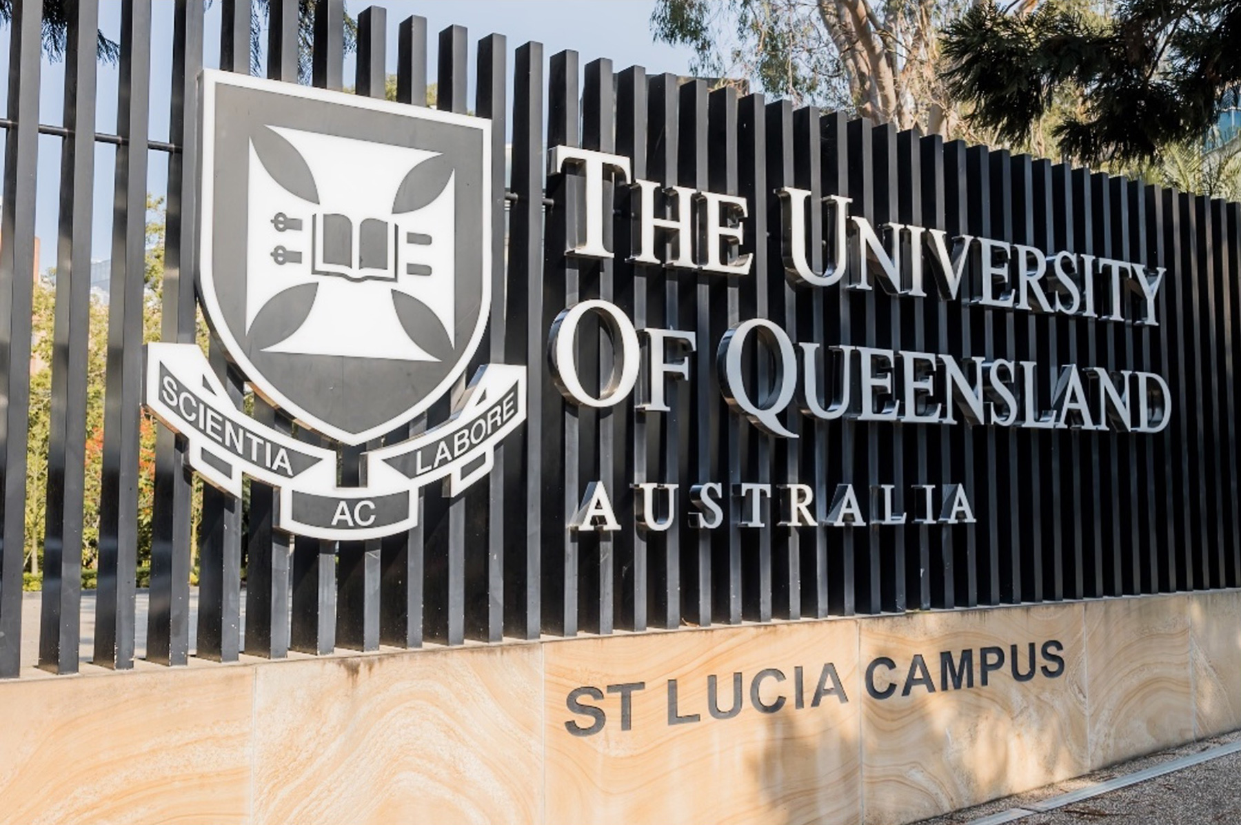
The University of Queensland, one of the Group of Eight Federal Universities in Australia, is known as a research-oriented university. It has achievements in biotechnology and nanotechnology fields. The Centre for Microscopy and Microanalysis (CMM) is the research infrastructure center supporting such cutting-edge research.
In July 2025, "Molecular Clamp technology" (*1) developed by the University of Queensland became a sensational topic. The license of this technology had been granted to Vicebio in the UK, an unlisted bio company headquartered in London, and the Vicebio was announced to be acquired by Sanofi S.A. , a leading French pharmaceutical company. The total amount of the initial payment reached 1.15 billion US dollars. The deal was the largest involving a company that was commercializing intellectual property from an Australian university.
Reference: Billion-dollar deal takes UQ vaccine tech to the world
Molecular Clamp technology makes it possible for us to respond as quickly as possible in vaccine production and will provide preparation against new pandemics and other virus infections in the future. What were the roles of CMM and the cryo electron microscope (cryo EM) in this research? We interviewed Prof. Roger Wepf, head of CMM, about the role of the center, and its future plan.
*1 Coronaviruses and influenza viruses infect their host through spike proteins on their
surfaces. These spike proteins are unstable and undergo structural changes (post-fusion form)
when triggered by the events such as binding to a receptor. In order to combat a viral
infection, vaccines based on the prefusion conformation, rather than the post-fusion
conformation, are more effective.
Molecular clamp technology is a method that stabilizes the spike protein in its prefusion state.
For example, the spike protein of SARS-CoV-2, the virus that causes COVID-19, can remain stable
by forming a trimer, in which three spike proteins are held together by a developed molecular
clamp (polypeptide).
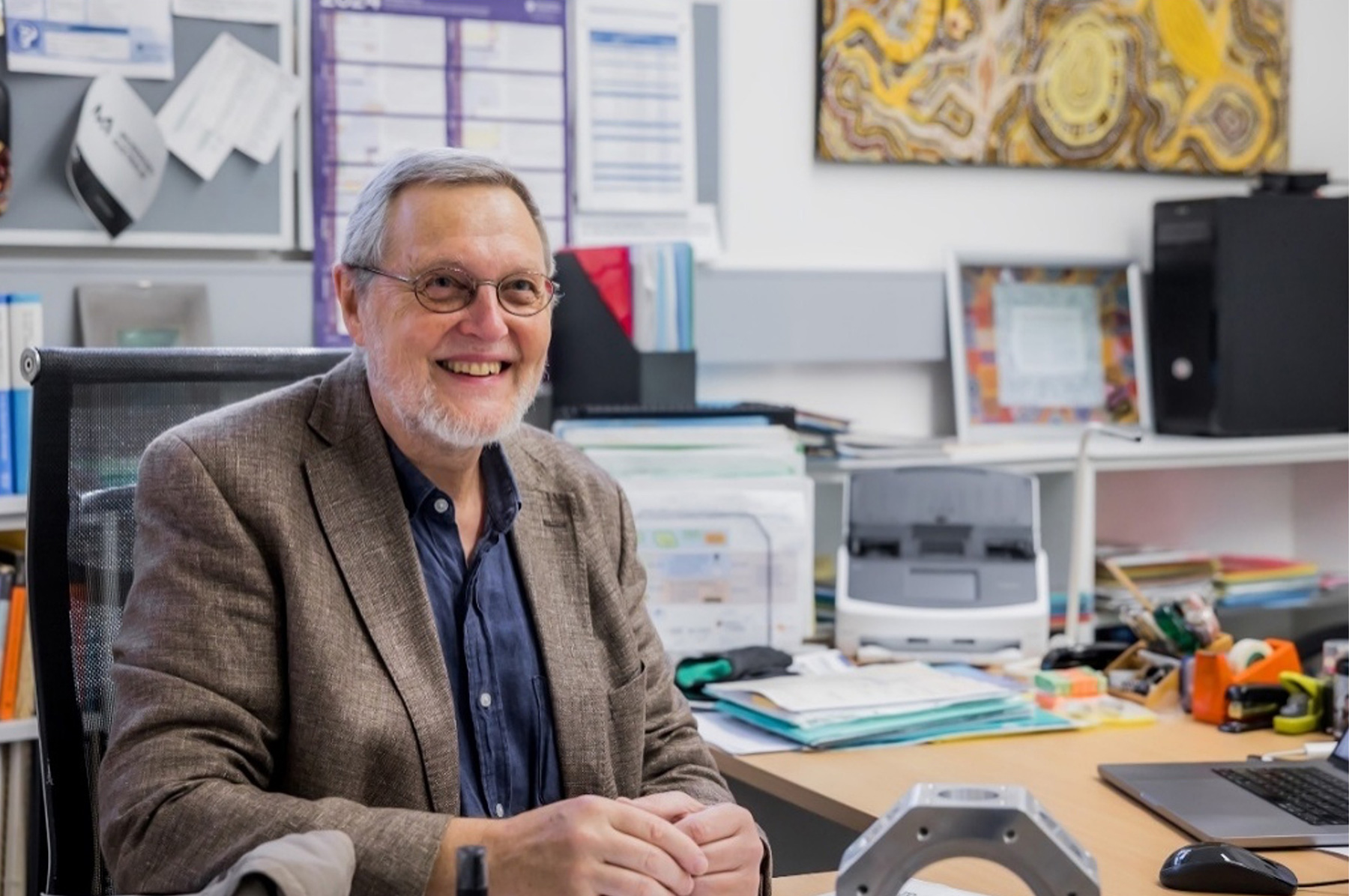
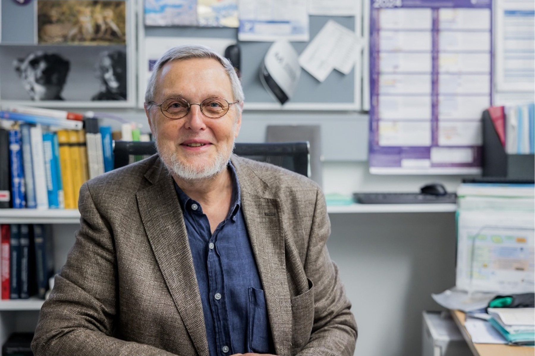
Prof. Roger Wepf, head of CMM
Q: What contribution did CMM and the cryo-EM make during the research process of Molecular Clamp technology?
A: Firstly, I would like to make it clear that CMM did not lead the
research of Molecular Clamp.
We supported the team of Paul Young Professor Emeritus, Prof. Daniel Watterson, and Prof. Keith
Chappell.
Reference: UQ vaccine tech strikes billion-dollar deal
Then, how did we support them? The spike protein that they were focusing on, changed its surface
structure, and CryoTEM single particle imaging helped them observe the different structures of
spike proteins.
The clamp as it names says is to clamp three spike proteins and make them into a stable trimer
as they usually are on SARS and other virus surfaces. By taking this form of a trimer, the solid
structure of spike proteins can be stabilized. This is exactly as a fixing jig (clamp) of parts.
When this trimer is entered inside the body as immune proteins, the body induces an immune
reaction and generates antibodies as if it would be infected by the virus.
We purchased JEOL's cryo TEM, "CRYO ARM™ 300" in 2019/20. At around the same time, we also purchased "CRYO ARM™ 200". Does Year 2020 ring a bell? Yes, the COVID-19 coronavirus disease outbreak occurred, just in time for the vaccine group after the installation of the new cryo TEMs they could go to work with structure determination. Therefore, these cryo TEM's quickly became powerful tools for the team developing the Clamp technology. They supported the earliest SARS-CoV-2 vaccine development research and subsequently the development of today's clamp technology.
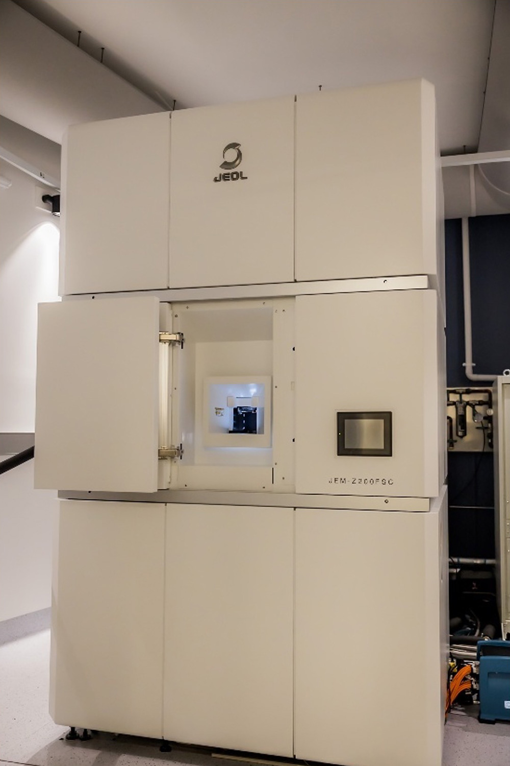
CRYO ARM™ 200
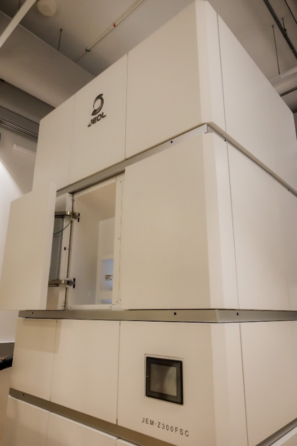
CRYO ARM™ 300
Q: Can you tell us about CMM again. What role is it playing and in what facility?
A: CMM is a significantly large scale research collaborative platform. We call it a CRP (collaborative research platform) focusing on state-of-the art electron, mass spectrometry and Xray characterization and imaging capabilities for man-made and natural materials. Basically, it underpins all experimental research fields of the university, from engineering, material science, life science, biology, quantum physics and devices and polymer complex materials. We support 700 to 800 projects in a year with around 750 users.
Unlike standard optical microscopes, most instruments require a high level of expertise to operate. We make sure that users are able to access and work with such instruments by engaging with excellent staff and teams. Supporting them so they can excel and carry out their research. Democratizing access to these frontiers technologies that is the role of the CMM.
Q: Is there anything you are keeping in mind as the head of the center?
A: I always hope to make various tools available for anyone. Here the expertise of staff becomes essential and CMM team members are working together so that cryo-TEMs or any other instruments can be utilized as easily as possible.
We also need to think into the future by planning ahead what researchers would like to use and develop a replacement and upgrade plan. We would like to update the instruments every few years to the latest models but this is not always possible - so clever upgrades help us to extend their live time or allow for unique new capabilities. For this purpose, we constantly need to apply for research funds and develop excellent ideas to enable the renewal of the platform. For me this is what I call technology scouting and local R&D, the most exciting part of my job.
Right now, we are examining whether we can provide new capabilities that are not available with the existing microscopic center. To be concrete, a workflow which we should call "cryo integrated" workflow is under examination. This is an idea to integrate different characterisation methods in a continuous series of workflow, including not only cryo SEM (scanning electron microscope), cryo TEM (transmission electron microscope), but also optical microscope and imaging mass spectrometry imaging etc with increased grade of automation. I believe that one of my major roles in CMM is to bring such idea forward into reality.
Normally, it is difficult to transfer a sample from one instrument to another. Our goal is to understand a sample from a comprehensive point of view. Which means, to investigate the sample at different scales, and to understand the sample by using more than one selection of chemical information, and various kinds of spectroscopic data.
Concretely, a low temperature transfer system, and a system that can identify the same region of the sample
at different scales are the ones we need to develop alongside research projects. What we are trying to
establish is, so to speak, an imaging pipeline across characterization modalities and instruments.
This is not because we simply would like to do it. Actually, we will be able to provide unique measurement
results to researchers of various projects faster while keeping a sample pristine (less artefacts).
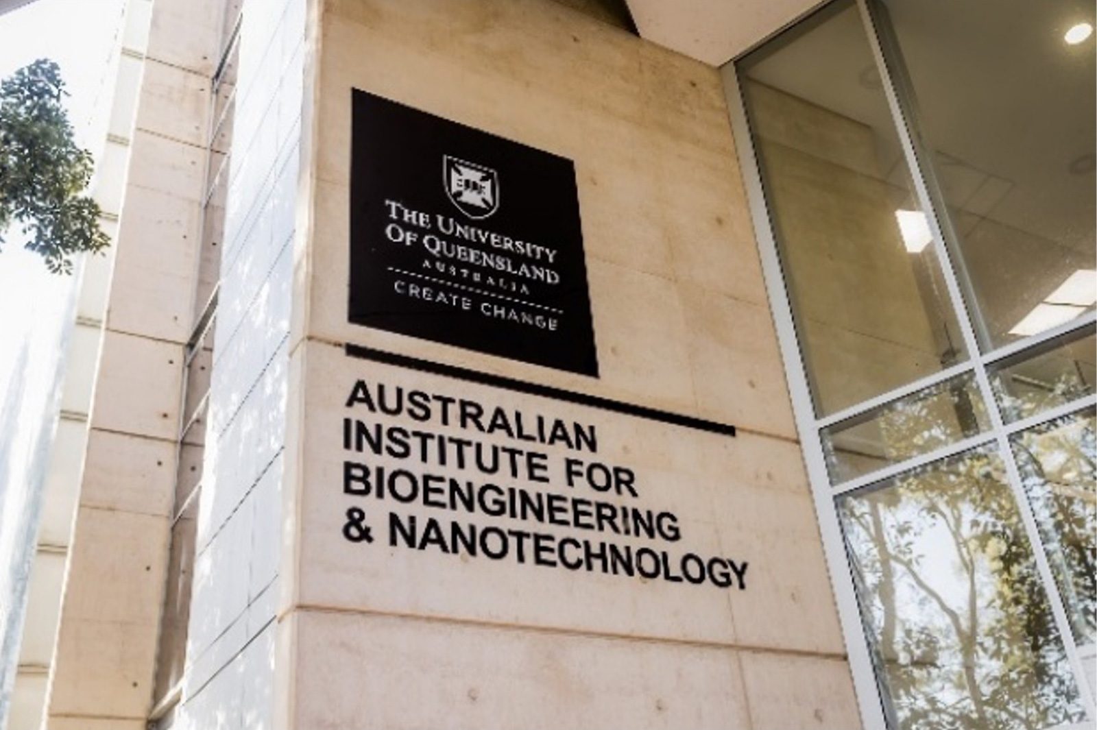
AIBN:Australian Institute for Bioengineering and Nanotechnology
In the facility, cutting-edge facilities such as CMM and excellent research center are consolidated.
Q: Please tell us why you chose to introduce a cryo TEM of JEOL Ltd.
A: We used to use a cryo TEM of another manufacturer. However, we had to perform every step by hand such as sample introduction and sample management. During procurement this time, two manufacturers were considered. We actually visited the laboratories of each manufacturer and tested their instruments with our own samples.
I used SEM and STEM with a cold field-emission electron gun when I was doing my doctoral degree. So, I knew that this type of electron gun could bring higher resolution, coherence and excellent phase contrast transfer(*2). I was very much concerned about working principles of JEOL's cold field-emission TEM. Therefore, we tested instruments at several locations including Tokyo, and examined instruments of JEOL's competitors, as well. As a result, we were able to confirm that JEOL's instruments could achieve far better S/N ratio and high resolution in the acquired images.
*2 Phase contrast transfer function:
The phase change of the electron wave cannot be observed (detected) in the TEM image normally. By converting
the phase change into an amplitude change, the phase change can be observed in the TEM image. The function
expressing what extent the amplitudes converted from the phase changes of the diffracted waves contribute
(are transferred) to the TEM image is called the phase contrast transfer function.
In addition, JEOL products have the omega filter (*3) included in the column. When a microscopic sample is handled, sometimes, so-called zero loss filtering (*4) can be required. This filter is integrated into the electron optical system. So, zero loss filtering can be easily performed as required as well a energy filtered image without the need of separate calculation or operation by installing an additional energy filter of a third party.
*3 Omega filter: An in-column type energy filter installed between the intermediate and projector lenses in a transmission electron microscope. It is mainly used to obtain filtered images (zero-loss images), zero loss CBED patterns, and energy-loss images.
*4 zerol loss filtering: Selection or removal of electrons that have lost a specific amount of energy when passing through the sample. This is used to improve the image contrast and enable elemental analysis.
JEOL instruments can hold up to 12 samples in a cryo port and 1 to 4 samples can be exchanged at one time. On the other hand, competitor's products needed the whole cartridge, all of 12 samples unloaded. Because of these main reasons and some others, we have selected JEOL products.
Q: I guess that you have had a severe situation under the COVID-10 pandemic such as travel bans and shutdowns of transportation networks. How did you find the support of JEOL?
A: The introduction of the cryo TEM proceeded smoothly. Right before the entry of COVID-19 in the country, the instruments were delivered and installations started. It was fortunate. However, it is not that there were no problems. We understand well that these instruments can always cause many problems in tuning and so on. However, we were able to devote ourselves to the original works. And then, we could contribute to the development of the Molecular Clamp. Frankly speaking, JEOL's support was extremely well and helpful to get started.
Q: How do you think the cryo EM will advance in the coming 5 to 10 years?
A: One thing is the size of biomolecule that can be handled. The combination of cryo EM and the single particle analysis method is now a new standard for structure determination. Moreover, molecule of 40,000Da (*5) or smaller will become the target. It is only a matter of time I guess.
By making the column where the electron gun is contained more stable, increasing the resolution of the camera, improving the processing speed and other areas, then, you are probably likely to achieve 20,000 Da. This combined with a high precision goniometer will also benefit cryo TEM Tomography and MicroED.
*5 Da (Dalton): Unit of mass where one-twelfth (1/12) of the mass of a carbon-12 atom is taken as one unit.
The other thing is the development of the next generation cryo TEM based on a cold field emission electron gun. This is the type of model with a relatively low accelerating voltage of 80 kv to 120 kv, high speed detector, and auto loader provided. This is what Richard Henderson (Nobel prize laureate in chemistry with cryo EM) describes as "democratized" cryo TEM. It is a low cost cryo-TEM that can be used by a wider range of people, which is the type he had long wished to develop.
*Reference: Nobel Prize in Chemistry 2017
In any case, for further development of such complex, deep tech instrumentation, I think it's inevitable for a company to proactively develop together with researchers across the globe next generations excellent product, which I know JEOL does well.
Profile
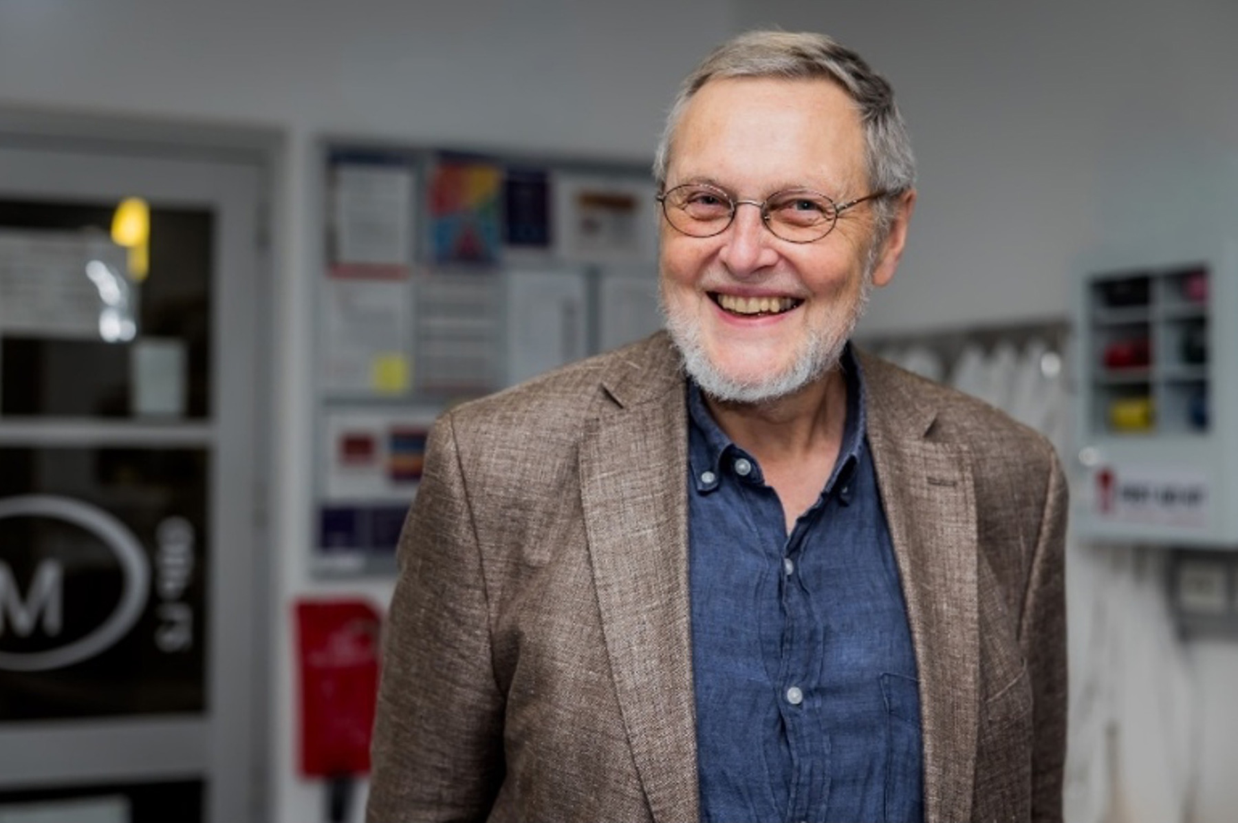
Prof. Roger Albert Wepf
Professor, Director of the Centre for Microscopy and Microanalysis (CMM), University of Queensland
Born in Strasbourg, France.
Roger had a degree in cell biology and structural research from the Swiss Federal Institute of Technology (ETH) in 1992. Following his postdoctoral research, he joined the physical instrumentation program in the
Max Haider Group at the European Molecular Biology Laboratory in Heidelberg where he engaged in the
development of cryo-electron microscopy technologies. Subsequently, he served as head of the analytical
microscopy department in a private sector in Hamburg.
In 2006, he became the director of electron microscope center, ETH Zurich. In 2014, he was appointed
Technical Director of the Center for Optical and Electron Microscopy (ScopeM). He served as the president of
the European Microscopy Society from 2012 to 2016.
This interview was conducted in November 2025. Professor Roger Wepf passed away on June 4, 2026. We are deeply grateful for his kindness, generous support, and willingness to share his insights in this interview. We extend our heartfelt condolences to his family, friends, and colleagues, and remember him with deep respect and gratitude. May he rest in peace.