


Ultra Flat Carbon BIG
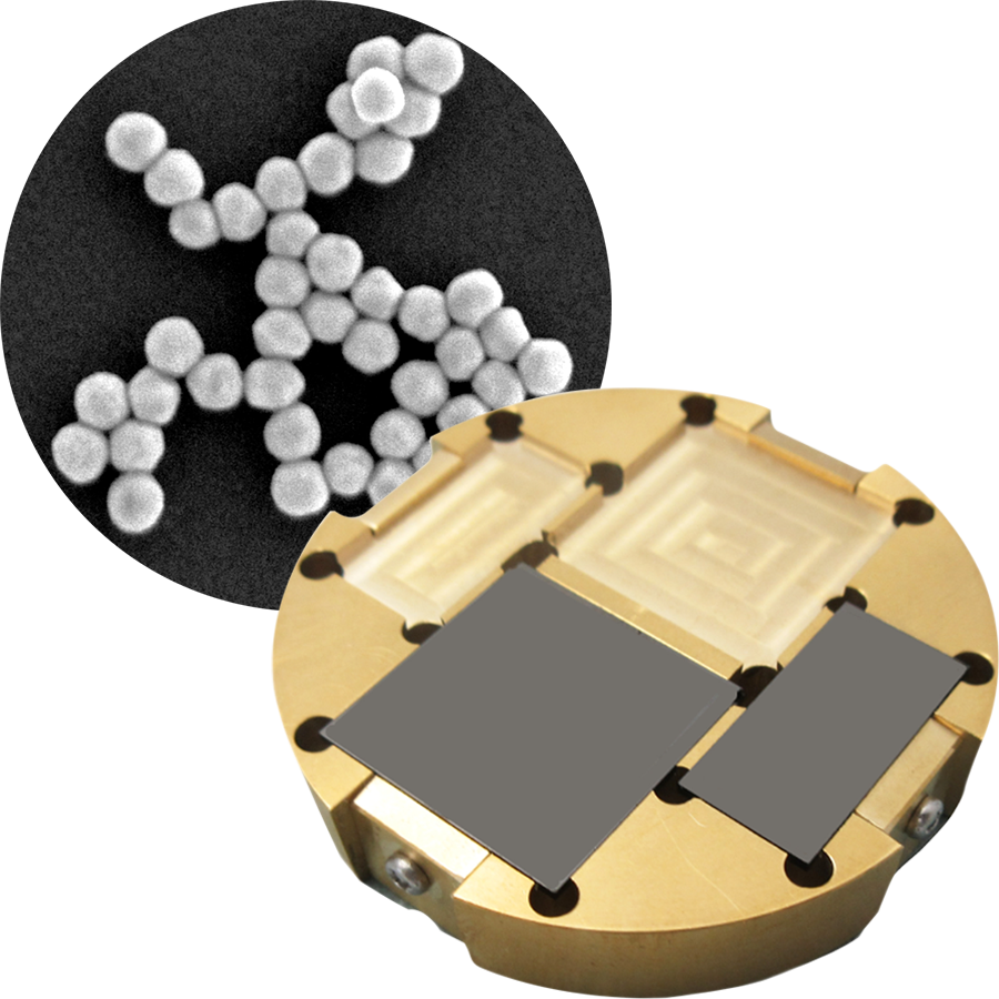
A groundbreaking new idea like never before!
SEM specimen stub ideal for observing ultrathin sections and ultrafine particles.
Features of Ultra Flat Carbon (Ultra Flat Carbon Stub*)
* Hereinafter referred to as UFC
Ultra Flat Surface
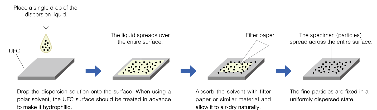
The solvent dispersion method is an effective technique for dispersing fine particles; however, surface irregularities on conventional specimen stubs can hinder proper particle distribution. In particular, dispersing nanoparticles requires a high degree of surface smoothness. Because the surface of UFC is ultra smooth, it enables uniform dispersion of specimens.
Ideal for Observing Low Contrast Particles Such as Organic Materials
The UFC is made of pure carbon, which reduces the signal generated from the specimen stub and allows observation without disturbing the specimen contrast.
Eliminates Issues Associated with Specimen Preparation Using Conventional Metal Grids
When using the UFC, there is no limit to the observation area for specimens such as biological specimens that continuously observe low magnification to high magnification, and the entire surface of the specimen can be observed.
Made of High Density Carbon—Ideal for Analysis
The UFC is conductive due to high density carbon. It does not interfere with observation and analysis like conventional silicon wafers and glass plates.
Procedure for the Solvent Dispersion Method
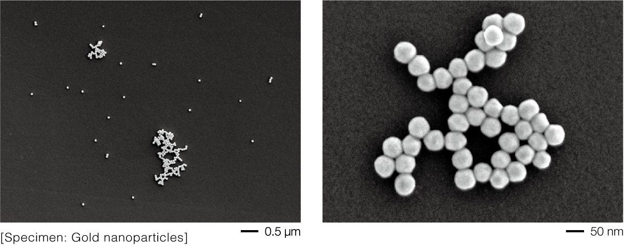
Also Ideal for Low Accelerating Voltage and High Magnification Observation
(SEM image; instrument: JSM-IT800, accelerating voltage: 2 kV)
Even at low accelerating voltage conditions, the background contrast is suppressed,
allowing the specimen contrast to be clearly and accurately represented.
Procedure for the Ultrathin Sectioning Method

Its large size makes it easy to place ultrathin sections, and multiple serial sections can be mounted, making it ideal for array tomography.
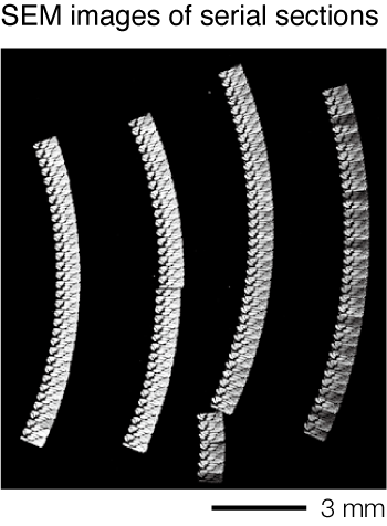
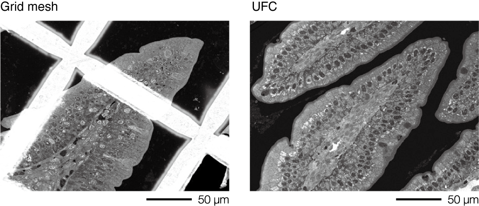
In a grid mesh, vignetting occurs in the field of view; however, with the UFC, there is no vignetting, allowing observation from low to high magnification. Any area of interest within the tissue section can be observed freely.
[Specimen: mouse small intestine]
Also Ideal for EDS Analysis
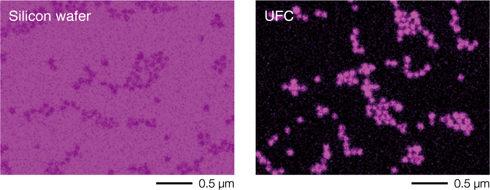
This is an example of silica particle analysis. When a silicon wafer is used as the substrate, it is difficult to obtain a clear Si-K EDS map; however, with the UFC, a sharp and well defined Si-K EDS map can be acquired.
Example of EDS analysis of ultrathin sections of Paramecium. EDS map of a phagoso
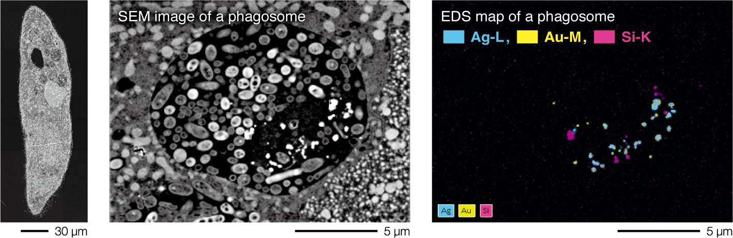
The distribution of nanoparticles (gold, silver, and silica) taken up into food vacuoles of Paramecium was visualized using EDS mapping.
* EDS detector: XFlash® 5060 FlatQUAD (Bruker Nano GmbH).
Product Introduction
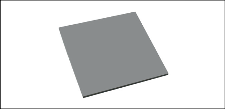
Ultra flat carbon BIG (L)
P/N 783160569
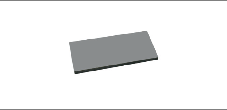
Ultra flat carbon BIG (S)
P/N 783160577
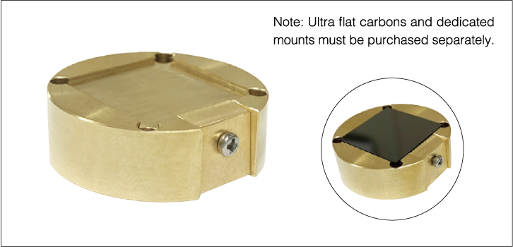
Ultra flat carbon BIG specimen mount
P/N 783163908
One Ultra flat carbon BIG (L), P/N 783160569, can be mounted, or two Ultra flat carbon BIG (S), P/N 783160577, can be mounted.
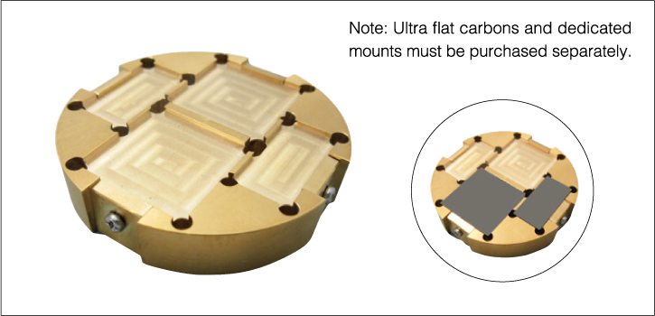
Ultra flat carbon BIG (plural number) specimen mount
P/N 783168128
Ultra flat carbon BIG (L) P/N 783160569,
Up to two Ultra flat carbon BIG (L), P/N 783160569,
and two Ultra flat carbon BIG (S), P/N 783160577, can be mounted;
or up to six Ultra flat carbon BIG (S), P/N 783160577, can be mounted.
Product Lineup
| P/N | Product Name | Specifications | Qty |
|---|---|---|---|
| 783160569 | Ultra flat carbon BIG (L) | 20 mm × 20 mm × 1 mm | 1 |
| 783160577 | Ultra flat carbon BIG (S) | 20 mm × 10 mm × 1 mm | 1 |
| 783163908 | Ultra flat carbon BIG specimen mount | φ32 × 10, vise type fastening | 1 |
| 783168128 | Ultra flat carbon BIG (plural number) specimen mount | φ51 × 10, vise type fastening | 1 |


Are you a medical professional or personnel engaged in medical care?
No
Please be reminded that these pages are not intended to provide the general public with information about the products.