


CRYO ARM™ 300S
Field Emission Cryo-Electron Microscope
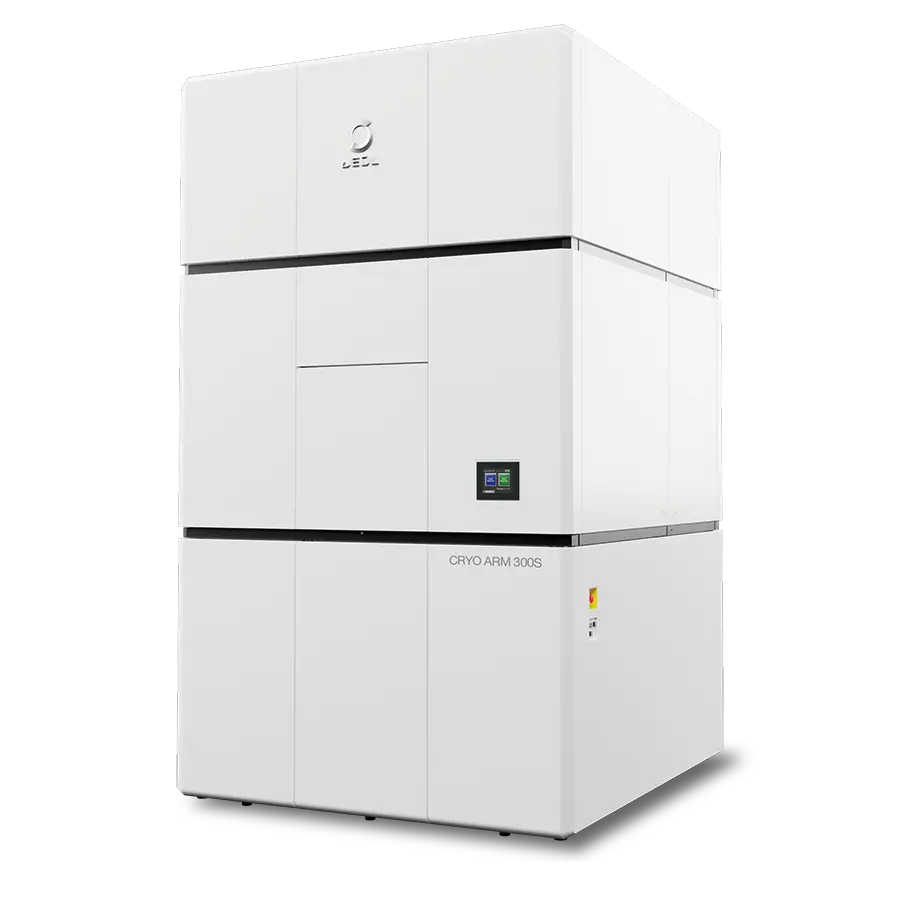
Welcome to the Ultra-High Resolution and Beautiful World
AI-driven automation and higher throughput make JEOL cryo-EM faster, more reproducible, and accessible to every operator. JEOL’s latest 300 kV cold field-emission cryo-electron microscope that offers single-particle analysis (SPA), cryo-electron tomography (cryo-ET), 3DED/MicroED and beyond, all in one highly automated instrument. The CRYO ARM™ 300S is engineered to deliver high-resolution structural data with the stability, throughput and ease of use that today’s structural-biology and drug-discovery laboratories require.
Features
CRYO ARM™ 300S × SmartScope
Single particle analysis data acquisition supported by AI
SmartScope 1 is an open-source software that supports data acquisition in single particle analysis. Using AI, it automatically detects areas suitable for imaging and can automate the entire process up to data acquisition. In addition, with an intuitive UI that organizes the operation flow in a simple manner and with necessary configurations kept to a minimum, even beginners can achieve stable data acquisition regardless of the level of experience.
1. Jonathan Bouvette et al., eLife, 11: e80047 (2022)
Workflow
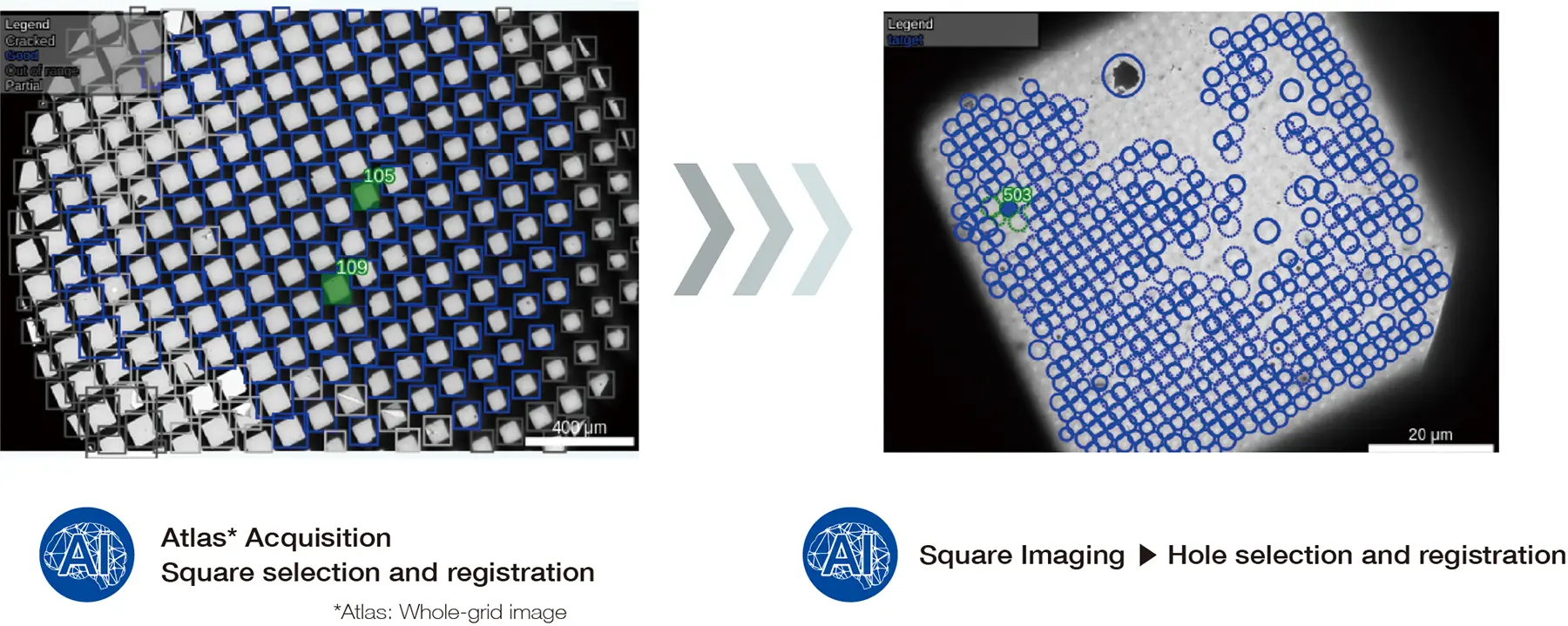

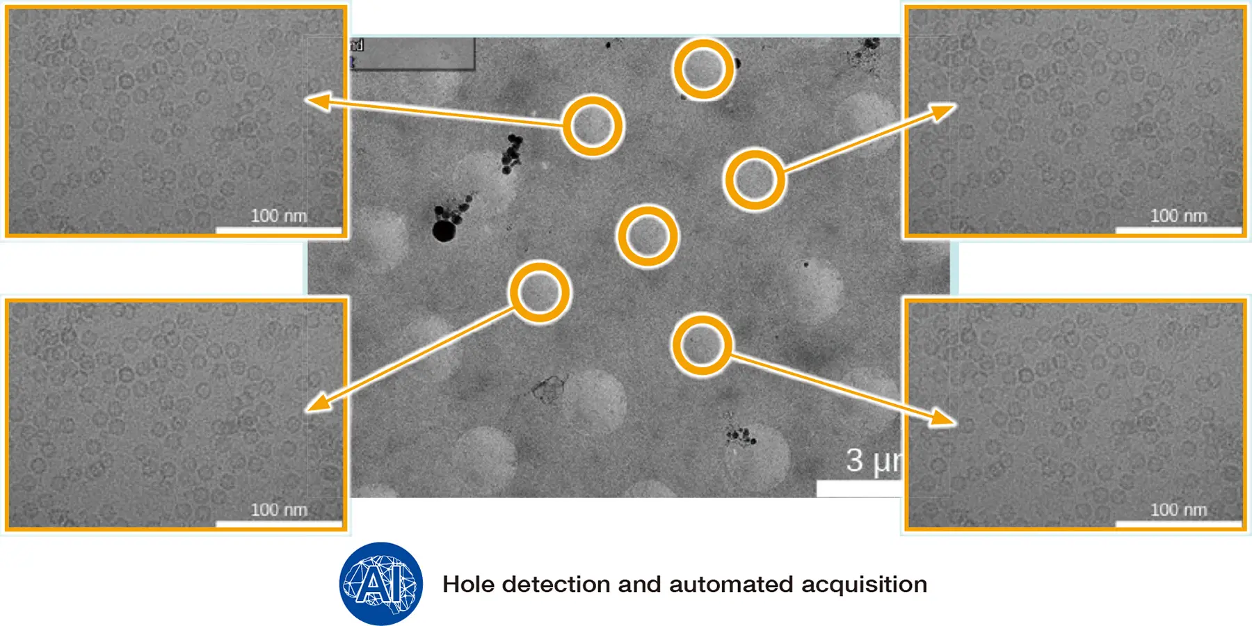
Interface
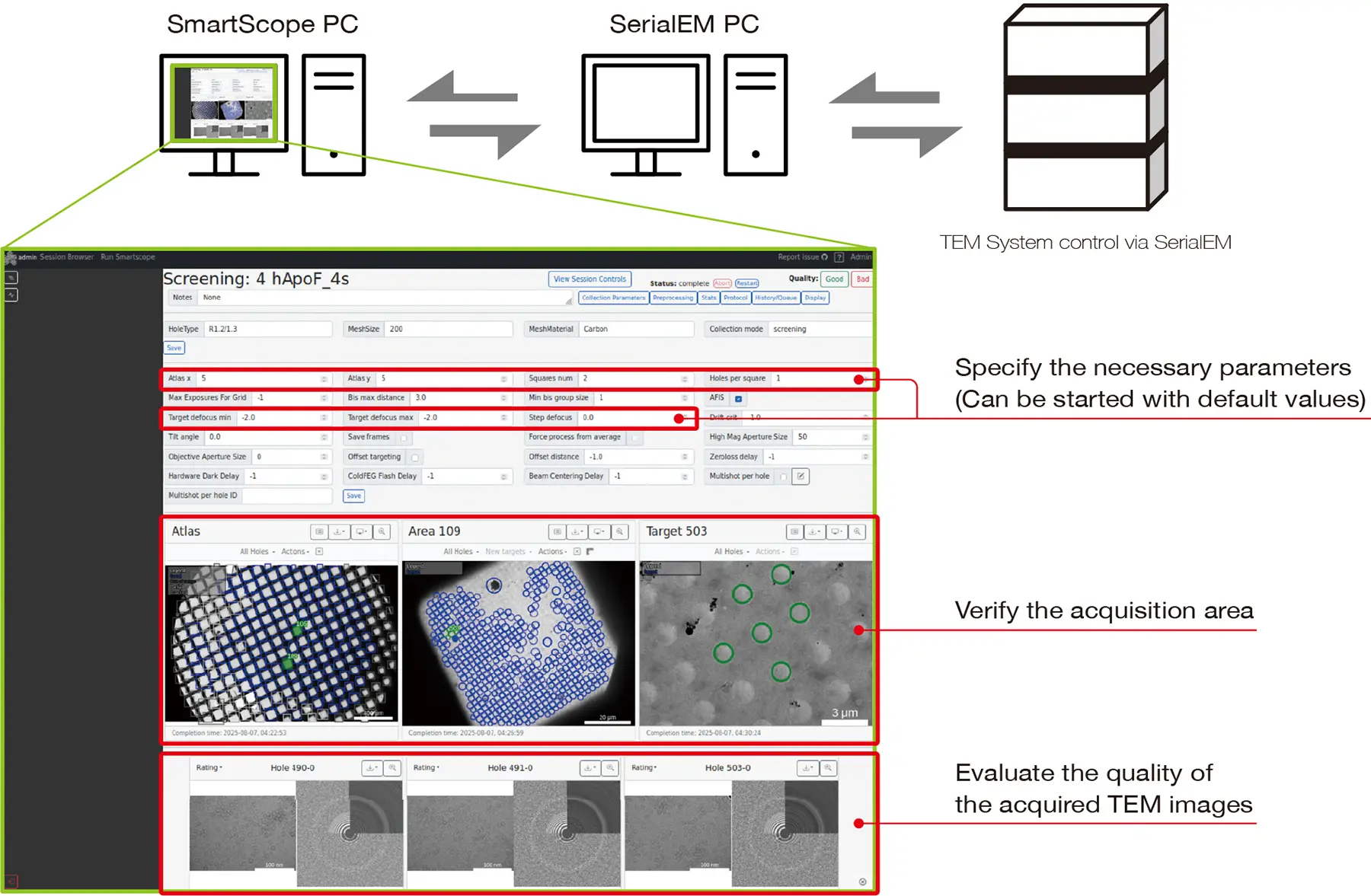
Result
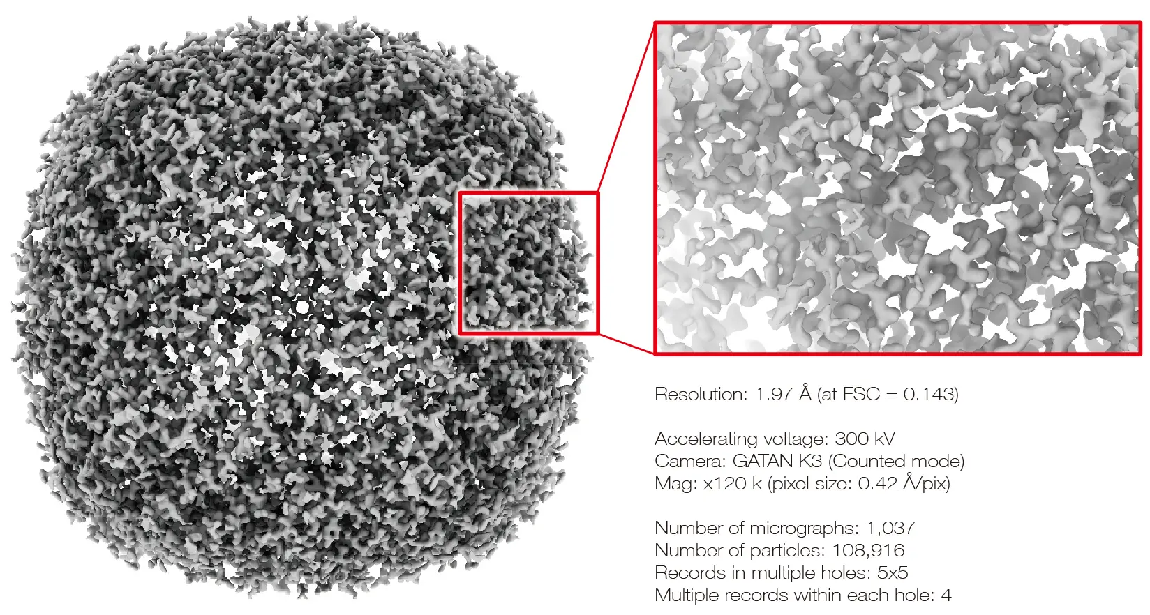
Apoferritin Structure with SmartScope
CRYO ARM™ 300S × System Innovations
High throughput, easy operation
Smart sample transfer system CRYO SPECPORTER™-S
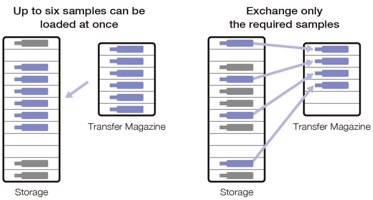
The advanced CRYO SPECPORTER™ -S separates the storage for sample preservation from the transfer magazines used for sample exchange. This permits selective handling of only the samples required during analysis, eliminating the need to load and unload all samples in the instrument during exchange. This supports stable and efficient operation, even in multiuser environments.
Enhanced cartridge handling with higher transfer capacity
The number of cartridges that can be loaded into a magazine at one time has expanded from four to six, reducing the frequency and time required for sample exchange, boosting throughput while maintaining stable sample storage environment.
"Endless" multi-grid imaging capability
A flexible sample transfer system allows selective, individual exchange of only the required samples. In multi-grid imaging, samples analyzed can be exchanged as needed while new samples are preloaded in advance, allowing uninterrupted overnight operation and maximizing data acquisition efficiency.
Sample storage in a clean environment
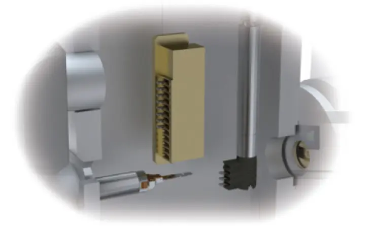
The storage is maintained under vacuum and at liquid nitrogen temperature, enabling sample storage for approximately one month in a clean, ice-accumulation-free state. This allows flexible sample management in accordance with analysis schedules.
Beam designed for maximum information yield
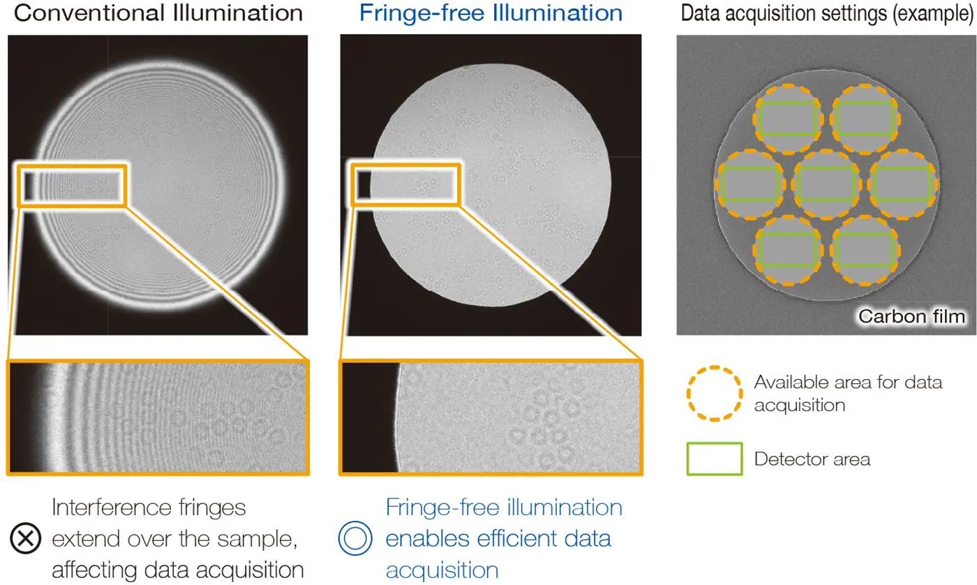
CRYO ARM™ 300S features fringe-free illumination, which prevents interference fringes during imaging. By making efficient use of the beam, data can be acquired with high throughput while minimizing damage to the sample.
Improved stability and flexibility of the electron beam
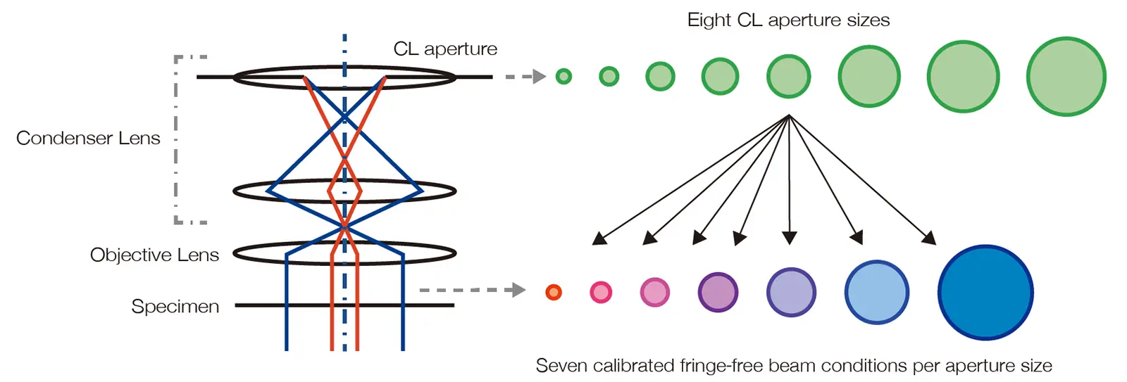
Improvements to the illumination system, including the condenser lens (CL) and CL aperture, refine the flexibility and stability of the electron beam. Enhanced lens demagnification ratio and aperture positioning accuracy improve beam positioning at the specimen plane. Eight aperture sizes and seven optimized fringe-free beam conditions for each aperture size enable ideal illumination across a wide range of specimens.
Longer consumable life for reduced maintenance
Roots pumps are used for their extended maintenance intervals, reducing downtime and ensuring high system uptime.
CRYO ARM™ 300S × CryoLameller
Cartridge-based cryo-CLEM workflow delivers high efficiency and high reproducibility
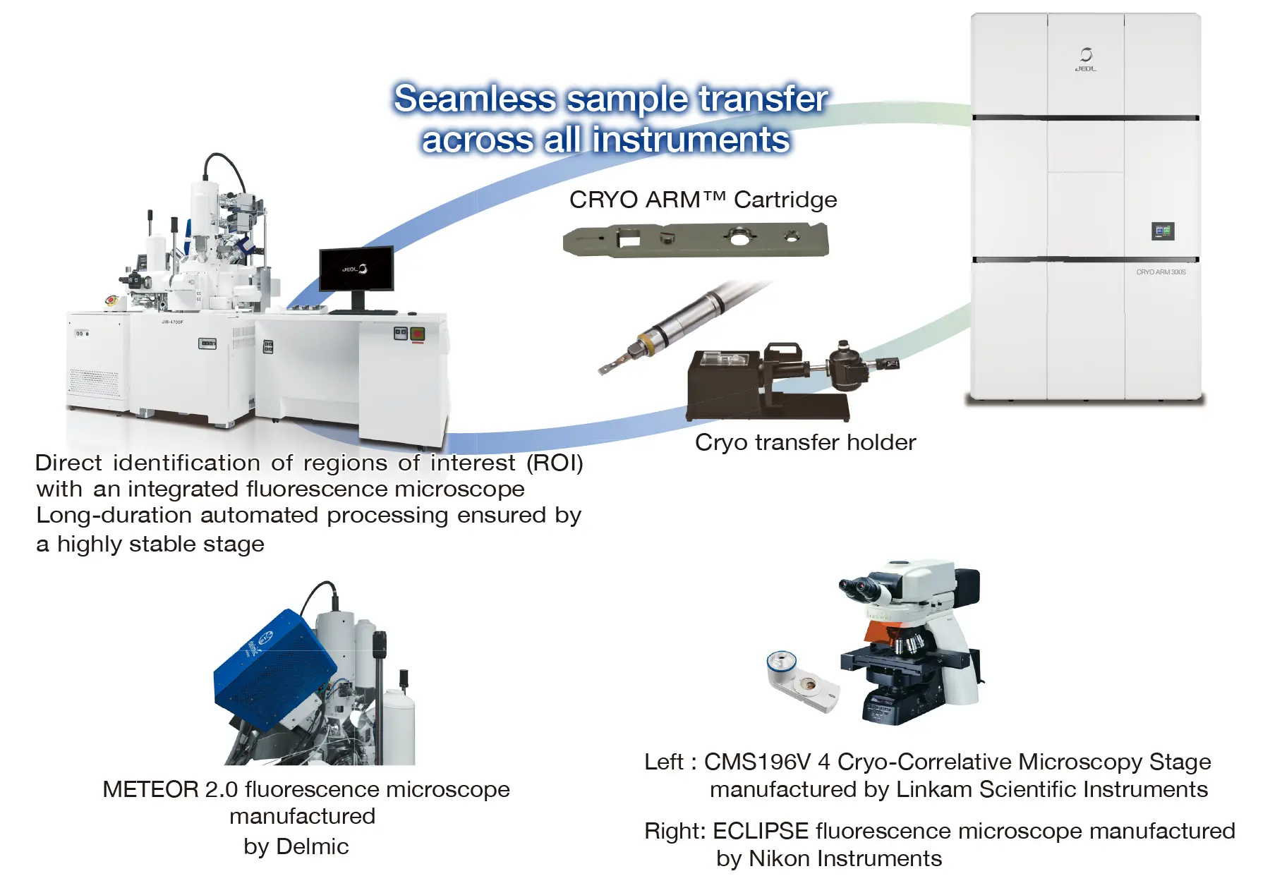
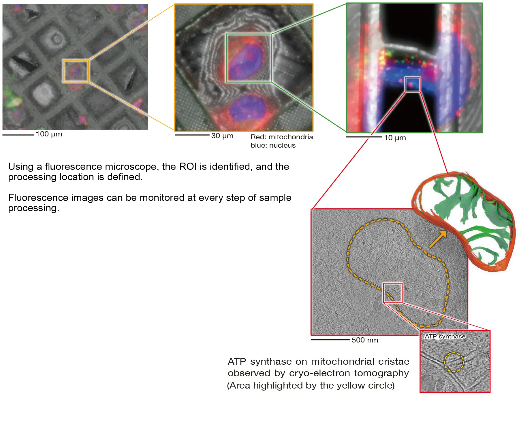
Integration with the CryoLameller (CRYO-FIB-SEM) enables a workflow that seamlessly connects sample preparation and analysis. From sample processing to data acquisition, the same cartridge holding the grid can be used throughout the workflow, allowing continuous operation while preserving positional information. With an integrated fluorescence microscope, intuitive and efficient area selection and positioning are achieved using correlative light and electron microscopy (CLEM).
CryoLameller – CRYO ARM™ Workflow
CLEM Application
CLEM imaging of fluorescently labeled mitochondria
Specifications
Main Instrument
| Electron Gun | Cold field-emission electron gun |
|---|---|
| Standard accelerating voltage | 300 kV |
| Energy Filter | In-column Omega energy filter |
Options
- Low accelerating voltage (Please make a separate inquiry about requests for other accelerating voltage except standard)
- STEM unit
- STEM detector (Bright-field/Dark-field)
- Camera system (Direct electron detector and related equipment)
- Smart Operation Software (AERO)
- EDM (Electrostatic Dose Modulator)
Specimen stage/Automated specimen exchange system / Specimen storage
| Specimen Stage | |
|---|---|
| Coolant | Liquid nitrogen, built-in automated liquid nitrogen filling system |
| Cooling temperature | 100 K or less |
| Specimen movements | |
| X, Y | Motor-driven (travel range: ±1 mm) |
| Z | Motor-driven (travel range: ±0.2 mm) |
| Tilt-X | Motor-driven (tilt range: ±70°) |
| Specimen exchange system | Air-lock, built-in automated cryotransfer system |
| Coolant | Liquid nitrogen, built-in automated liquid nitrogen filling system |
| Cooling temperature | 105 K or less |
| Specimen exchange cartridge | Up to 6 specimens can be exchanged at once. |
| Specimen storage | Capacity of up to 12 specimens |
Application
Gallery
CRYO ARM™ 300S × Applications
SPA・Tomography・3DED/MicroED in a single system
Single Particle Analysis
High-resolution three-dimensional structures can be determined by averaging images of numerous identical particles embedded in vitreous ice, even for samples that are difficult to crystallize
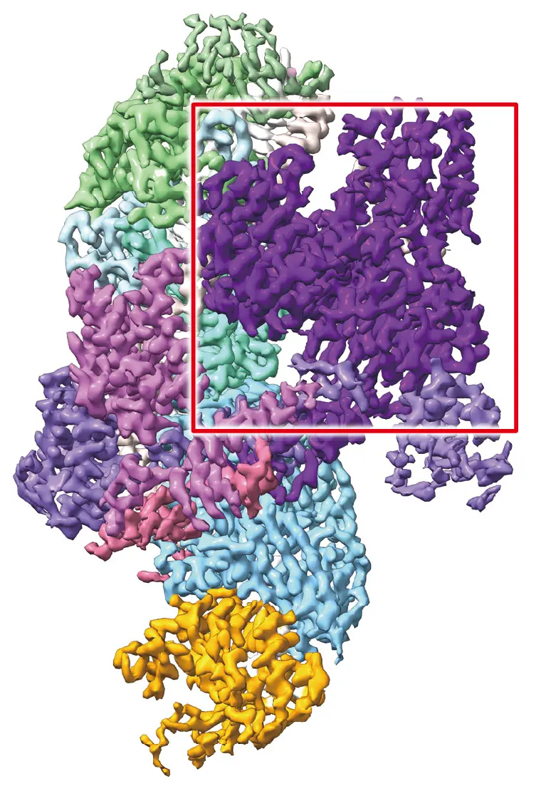
Resolution: 2.8-2.9 Å
Cas5-Cas-7-crRNA-CTR-Cas14 quinary complex
Data provided by Dr. Heng Zhang at Tianjin Medical Univ.
CRISPR-Cas system
CRISPR Cas is a genome editing system derived from an adaptive immune mechanism, in which crRNA from CRISPR loci works with Cas enzymes. Among its various types, the type VII system shown on the left is compact and offers the potential for highly precise genome editing. The CRYO ARM™ 300S enables high resolution structural determination, contributing to the elucidation of genome editing mechanisms.
Cas14 with site-specific cleavage activity Structural determination at atomic resolution has elucidated the cleavage mechanism of CTR in type VII CRISPR-Cas systems.
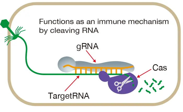
CRISPR-Cas Mechanism
hERG channel (human ether-a-go-go-related gene channel)
The hERG channel is a protein that forms a pathway for potassium ions across the membrane of cardiac muscle cells. Inhibition of its function can lead to life threatening arrhythmias. The figure below presents a high-resolution structure of the hERG channel bound to an anticancer drug, providing critical insights for predicting and mitigating cardiac side effect risks in drug development.
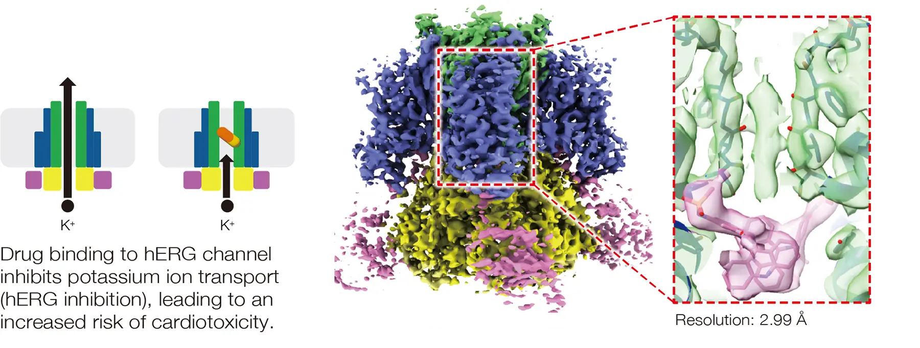
Anticancer Drug-Bound hERG channel
Data provided by Dr. Takeshi Murata and Ryuhi Yoshioka at Chiba Univ.
Tomography
Three-dimensional structures are visualized by reconstructing data acquired while tilting the specimen
Euglena
Euglena is a unicellular organism that exhibits characteristics of both plants and animals and is utilized in applications such as functional foods and biofuels. By observing lamellae prepared with the CryoLameller (CRYO-FIB-SEM) using the CRYO ARM™ 300S, cellular organelles and their structures can be clearly visualized.
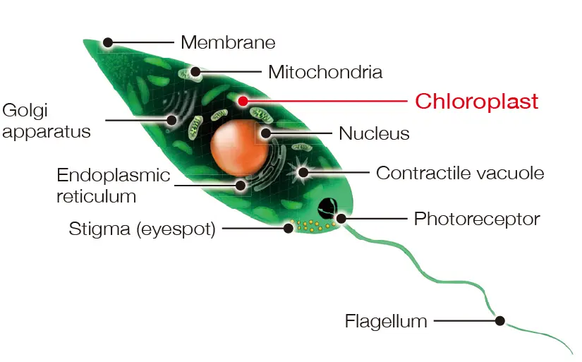
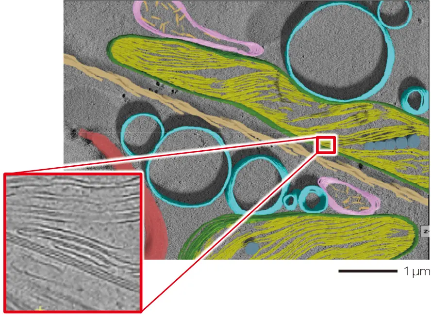
The double-membrane structure on the chloroplast surface and thylakoid membranes can be clearly visualized.
Euglena
Sample provided by Dr. Roland Fleck at King's College London
Lipid nanoparticle (LNP)
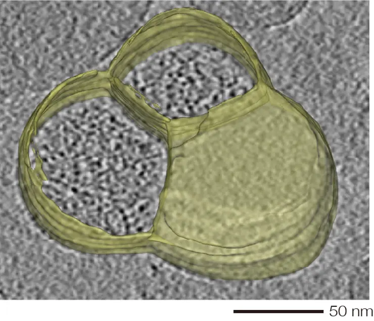
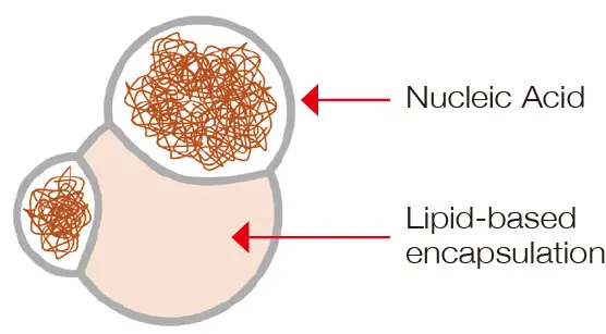
Visualization of nucleic acids encapsulated in lipid nanoparticles.
LNP including mRNA
Sample provided by Dr. Keisuke Ueda at Chiba Univ.
Lipid nanoparticles are key carriers in drug delivery systems (DDS) and have attracted significant attention as delivery technologies for nucleic acids such as siRNA and mRNA. The CRYO ARM™ 300S enables high resolution visualization of LNP internal structures and nucleic acid localization, offering in-depth analysis for DDS design and optimization.
3DED / MicroED
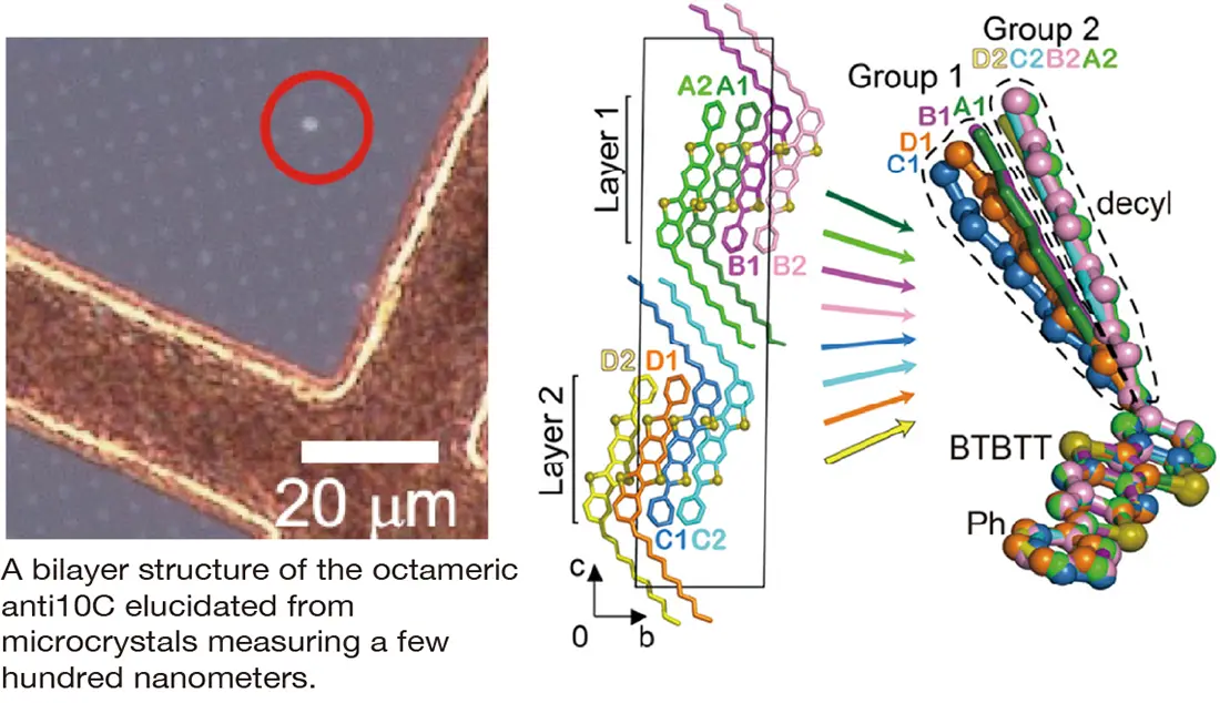
Atomic-resolution crystal structures are resolved from electron diffraction data obtained from microcrystals
Organic semiconductor
Organic semiconductors are used in applications such as OLED displays in smartphones and solar cells, combining excellent flexibility with processability. The figure on the right shows the structure of a material expected to be used in next-generation transistors, determined from microcrystals using 3DED / MicroED with reduced electron-beam damage.
Ph-anti-benzothieno[5,6-b]benzothieno[3,2-b]thiophene-C10 (antiC10)
Data provided by Dr. Kouji Yonekura at RIKEN, SPring-8
Center / IMRAM, Tohoku Univ.
Related Products
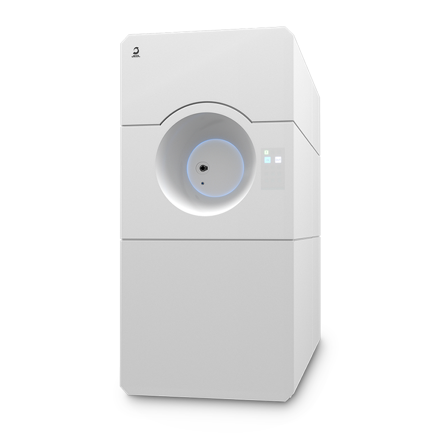
JEM-120i Electron Microscope
The JEM-120i is a newly developed Transmission Electron Microscope with a 120 kV accelerating voltage and is based on the concepts of “Compact”, “Easy To Use”, and “Expandable”. With the new external appearance, this instrument has evolved into a useful tool that anyone can use easily, from operation to maintenance.
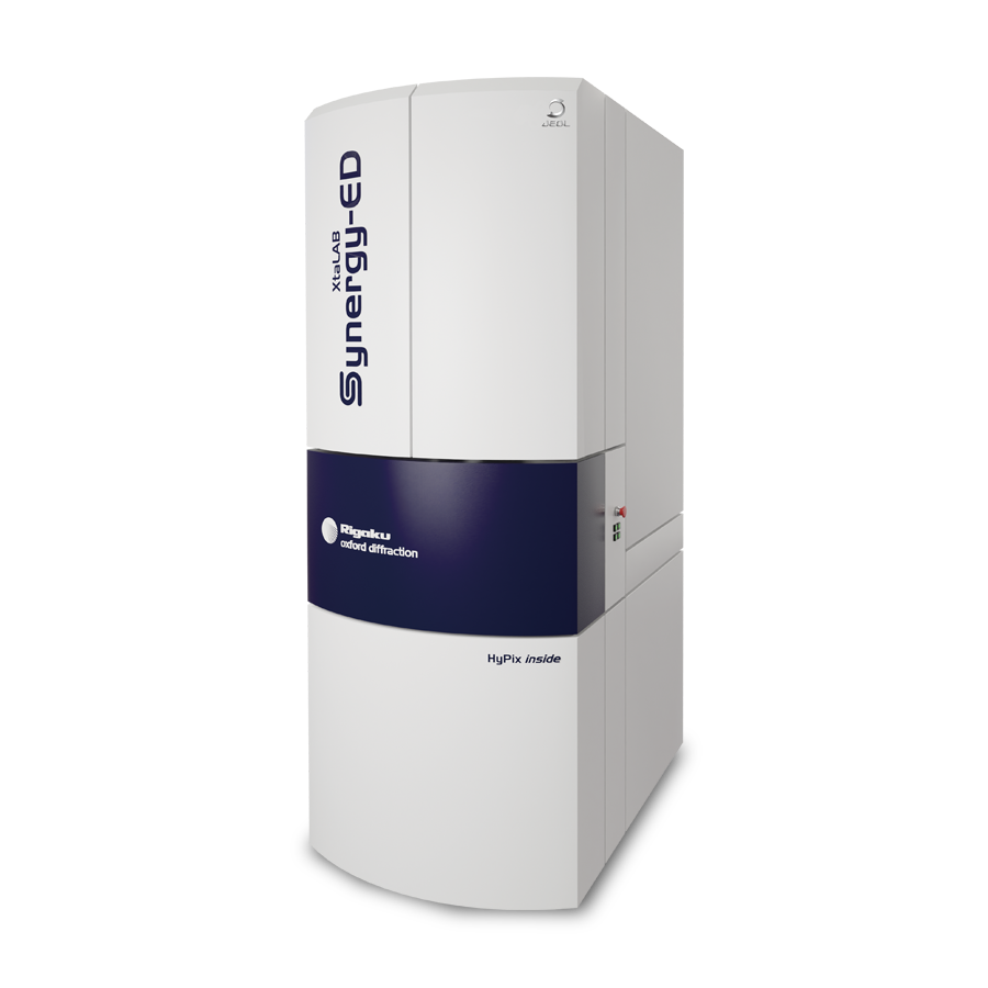
XtaLAB Synergy-ED Electron Diffractometer
XtaLAB Synergy-ED is a completely new electron diffractometer born from the synergy of Rigaku and JEOL's core technologies.
By integrating the flow from selection of measurement samples (nanocrystals) to data collection and analysis, electron diffraction structure analysis can be easily used by non-specialists who lack the expertise in electron microscopy and crystallography that is conventionally required.
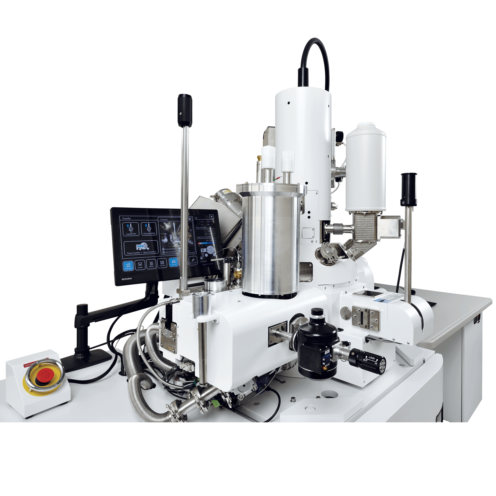
CRYO-FIB-SEM CryoLameller
This CRYO-FIB-SEM system incorporates a liquid nitrogen cooling stage and a cryocooled specimen transfer mechanism for frozen specimens, making it possible to prepare TEM specimens such as biopolymers. The specimen transfer mechanism has a built-in sputter coating function. Therefore, this CRYO-FIB-SEM system alone can perform a series of processes to create TEM specimens from frozen specimens including conductivity coating, protective film forming, and FIB processing. In addition, by using JEOL's CRYO ARM™ cartridge, direct specimen transfer to the CRYO ARM™ after TEM specimen preparation becomes easier.
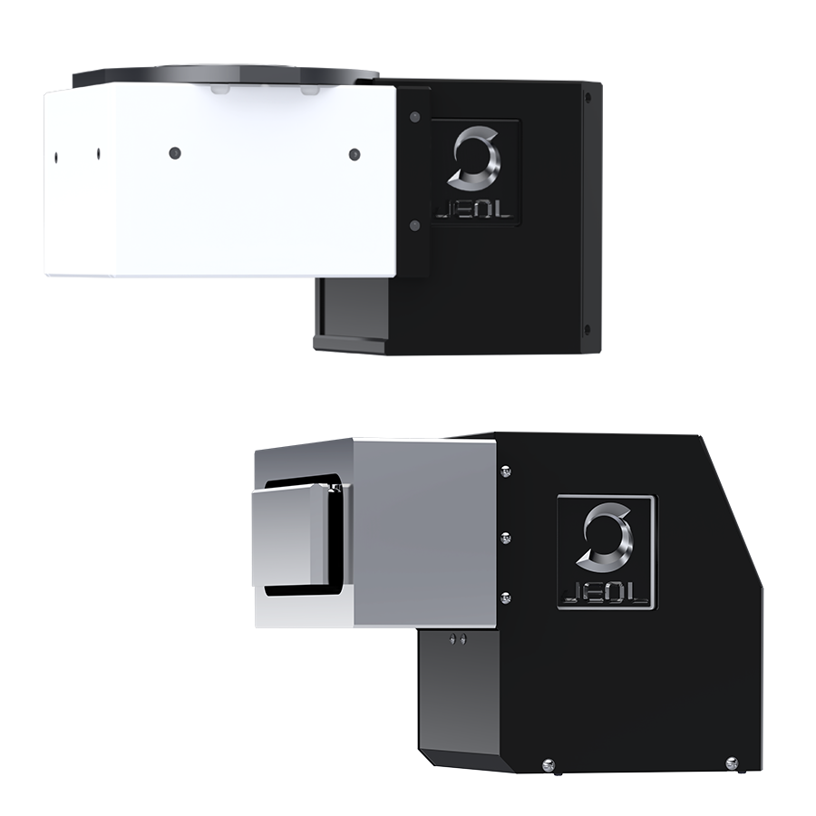
SightSKY™ Series High-Sensitivity, Low-Noise Fiber-Coupling CMOS Camera
High-sensitivity, low-noise 19 M pixel CMOS sensor enables imaging of fine specimen detail and obtains high signal to noise images even at low electron doses. A Global shutter and high frame rate (58 fps/full pixel mode) enable image series acquisitions with less artifacts during in-situ dynamic observation studies. The SightSKY™ camera system offers integration with JEOL’s FEMTUS™ Integrated Analysis Platform, providing user-friendly operation and data acquisition.
More Info


Are you a medical professional or personnel engaged in medical care?
No
Please be reminded that these pages are not intended to provide the general public with information about the products.