【DISCONTINUED】ClairScope Atmospheric Pressure Scanning Electron Microscope
DISCONTINUED
This product is no longer available.
If you would like to know the latest information about your preferred product or to find out more about alternatives, please click on the link below. We hope you will continue to use our products.
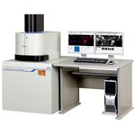
ClairScope is an instrument to observe specimens by using an Atmospheric Scanning Electron Microscope (ASEM) and an optical microscope. Since specimen remains at atmospheric pressure or in a solution during observation, the original and natural state can be captured.
Features
Native state (liquid) observation
ASEM imaging of specimens still steeped in liquid is possible, enabling imaging of biological materials without the need for skillful pre-treatment, such as dehydration/ desiccation, which also greatly decreases the preparation time
Concurrent imaging with an optical microscope
The Clairscope combines both an optical microscope and ASEM in one instrument. After observation with the optical microscope, ASEM imaging at the same location is possible with a click of the mouse.
Dynamic observation
Since the specimen chamber is open, it is possible to perform evaporation or introduce a reagent from outside. The reactions after instillation of a reagent or accompanying the evaporation can be observed with the ASEM in real-time.
Specifications
Scanning Electron Microscope (SEM)
| Resolution | 8 nm(30 kV) |
|---|---|
| Magnification | 100 × to 100,000 × |
| Operating conditions memory | Illumination system, specimen stage, etc. |
| Acceleration voltage | 10、20、30 kV |
| Filament | Factory pre-centered filament |
| Electron gun | Fully automatic, manually adjustable |
| Condenser lens | Zoom condenser lens |
| Objective lens aperture | 1 stage, with XY fine adjustment |
| Stigmator memory | Stigmator memory |
| Image shift | ±50 μm |
| Automatic functions | Focus, exposure, stigmator |
| Image memory(SEM) | 640×480 pixels, 1,280×960 pixels,2,560×1,920 pixels |
Optical Microscope (OM)
| Optical system | Infinity-correction optical system |
|---|---|
| Observation | Bright-field image, fluorescent image |
| IlluminatorHg lamp | Epi-illumination floodlight tube 100 W (lifetime: 300 h) |
| Focus | Electrically driven Stroke: 30 mm |
| Mirror unit | Fluorescent (U-MWU2), Bright-field (U-MBF3) ※ Up to 6 installable (optional) |
| ND filter | 6, 12, 25, 50, 100%, electrically switched |
| Objective lens | No-cover liquid-immersion lens, 40× |
| CCD camera | High-sensitivity color CCD 1,600×1,200 pixels |
| Automatic functions | Exposure, white balance |
Specimen holder
| Film-windowed dish | 35 mm diameter, window 0.25 mm square角 |
|---|---|
| Liquid capacity | Up to 3 cc |
| Specimen stage | XY ±2.5 mm, electrically driven |
Other
| Computer | IBM PC/AT compatible |
|---|---|
| OS | Windows® Vista * |
| Viewing monitor | 19 inch LCD ×2 |
| Multiple-image display | 2 images, 4 images |
| Measurement | Provided |
| Image format | BMP, TIFF, JPEG |
| Automatic image save | Provided |
| Evacuation system | Fully automatic, TMP ×1, RP ×1 |
| Eco/standby mode | Vacuum shutdown, display shutdown |
| Safety devices | For power failure, vacuum degradation, thin-film damage |
*Windows Vista is a registered trademark of Microsoft Corp.
Application
Application JASM-6200
More Info
Are you a medical professional or personnel engaged in medical care?
No
Please be reminded that these pages are not intended to provide the general public with information about the products.