miXcroscopy™
Linked Optical & Scanning
Electron Microscopy System
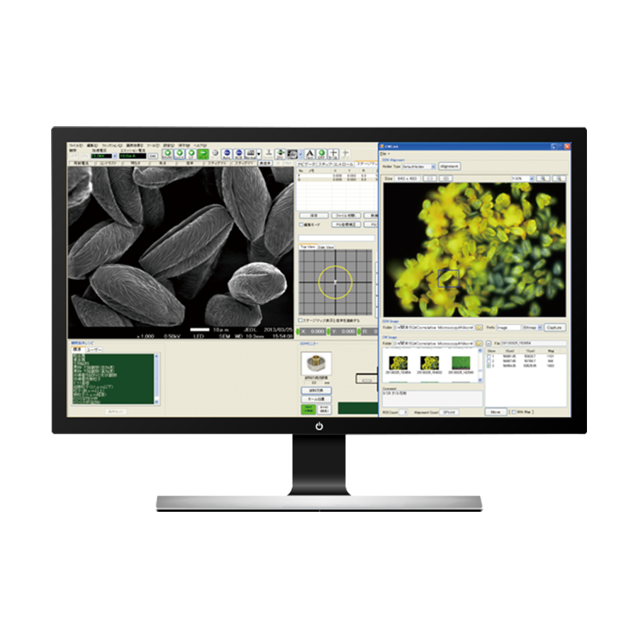
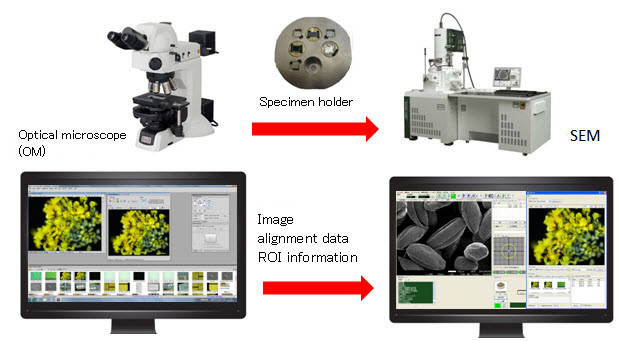
The same specimen holder can now be used for both the optical microscope and the scanning electron microscope. As a result, by managing the stage information with dedicated software, it is possible for the system to record the locations observed with the optical microscope, and then further magnify the same areas with the scanning electron microscope to observe the fine structures at higher magnification & higher resolution.The observation targets found with the optical microscope can be seamlessly observed with the scanning electron microscope without having to search for the target again. It is now possible to smoothly and easily compare and verify the optical microscope images and scanning electron microscope images.
Features
System Outline
Applicable models:
Field Emission Scanning Electron Microscope
Electron Probe Microanalyzer (EPMA)
Data acquisition and intuitive observation with the use of color
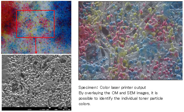
By adding visible light color information from the optical microscope image (which cannot be obtained with the SEM image) it provides an SEM image with a more intuitive visual effect.
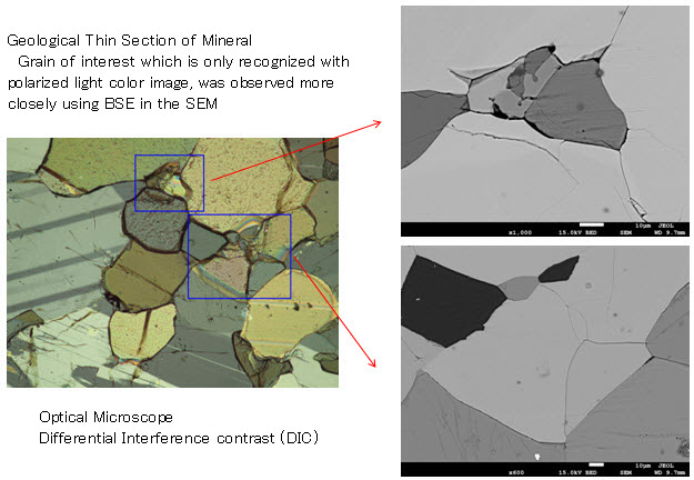
Smooth target search takes advantage of the features of the optical microscope
Prevents damage to the specimen from the electron beam
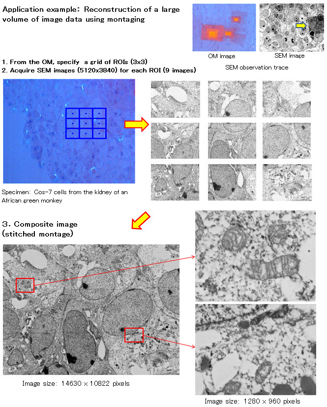
Application
Application miXcroscopy
Related Products
Scanning Electron Microscope (SEM)
Electron Probe Microanalyzer (EPMA)
More Info
Are you a medical professional or personnel engaged in medical care?
No
Please be reminded that these pages are not intended to provide the general public with information about the products.