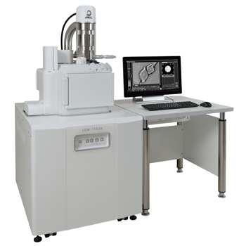
【DISCONTINUED】JSM-IT500 InTouchScope™ Scanning Electron Microscope
DISCONTINUED
This product is no longer available.
If you would like to know the latest information about your preferred product or to find out more about alternatives, please click on the link below. We hope you will continue to use our products.
The JSM-IT500 is a new model of JEOL InTouchScope™ series.
Equipped with our sophisticated Analytical series, the JSM-IT500 facilitates any analyses from specimen loading to report generation.
Features
Video introducing the JSM-IT500
◆Clicking the Play button starts the video (approx. 2 minutes).
◆ Features and functions of the JSM-IT500 InTouchScope™ are introduced.
3 key points Fast and Easy Analysis!
①
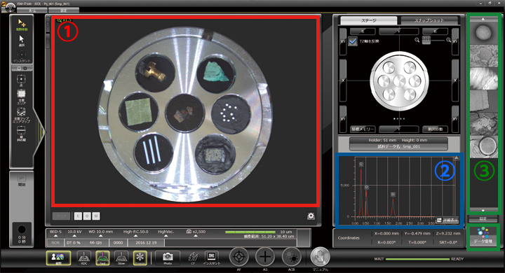
Key point 1 : Zeromag
The displayed Holder Graphics or CCD image enables you to locate the specimen area or specify analysis positions. You can rapidly search the specimen area and specify positions for multiple-field serial analysis.
②
Key point 2 : Live Analysis (Only for A/LA)
With "Live Analysis", the embedded EDS system shows a real time EDS spectrum during image observation. Elemental composition or "Alert" for element of interest is displayed on a live image.
③
Key point 3 : Integrated data management software
This user-friendly software enables you to select and review SEM images and analysis results through the data management area. You can also generate a report with a single click.
Key point 1 : Zeromag for True Integration of Optical and SEM imaging
Smooth operation up to high magnification observation!
"Zeromag" facilitates navigation with seamless transitioning from the optical CCD image to SEM image.
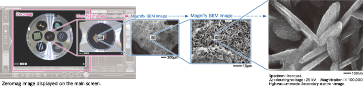
With Zeromag, automated simple large-area observation and EDS analysis can be made.
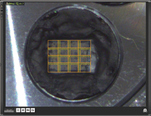
Montage setup with Zeromag.
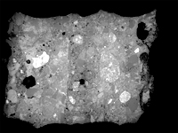
Montage result: 4 x 4
(Left: Backscattered electron composition image, Right: Ca map)
Specimen: Concrete. Accelerating voltage: 15 kV
High-vacuum mode. Area: Approx. 4 mm x 3 mm
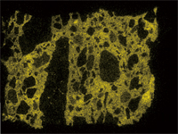
With Zeromag, it is easy to set up one or more montage areas for imaging and analysis. “Montage” function is effective to acquire detailed information for identifying foreign materials over large areas.
Key point 2 : Live Analysis - Seamless SEM and EDS -
With our analytic series (Live Analysis), the EDS system shows a real time EDS spectrum during image observation. You will easily find elements of interest and unexpected elements.
①
Element
The main elements existing in the measurement area are displayed. You can display “Alert” by specifying an element.
②
Spectrum
The characteristic X-ray spectrum from the analysis area and automatic qualitative analysis results are always displayed.
③
Single-click enables you to switch between the Electron microscope operation screen and analysis detail display screen.
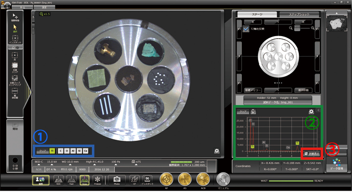
Key point 3 : Integrated data management software SMILE VIEW™ Lab for Seamless report generation
SMILE VIEW™ Lab is a fully integrated data management software which links the CCD image, SEM images, EDS analysis results and corresponding stage coordinates for fast report generation or re-call of specimen position for further study.
①
Names of each field are displayed.
②
Data search is enabled from specimen name, creation time, data type, etc.
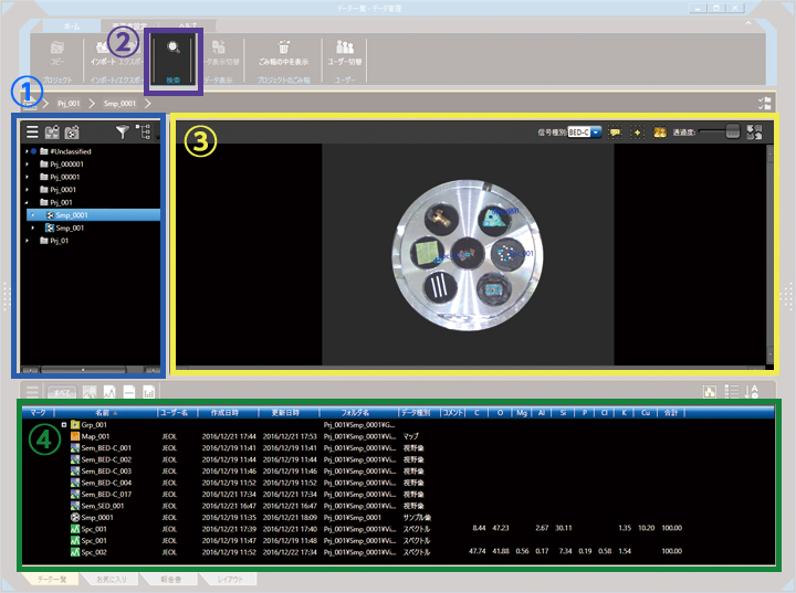
③
Positions of each field are displayed on Holder Graphics or CCD image.
④
Data is displayed in list form, which includes analysis data, quantitative analysis results of elemental map, spectra, etc., in the selected fields.
Specifications
| Resolution | High vacuum mode: 3.0 nm (30 kV) 15.0 nm (1.0 kV) |
|---|---|
| Low vacuum mode※1: 4.0 nm (30 kV BED) | |
| Direct magnification | x 5 to x 300,000
(Defined with a display size of 128 mm x 96 mm) |
| Displayed magnification | × 14 to × 839,724 (on the monitor)
(Defied with a display size of 358 mm x 269 mm) |
| Electron gun | W filament, Fully automatic gun alignment |
| Accelerating voltage | 0.3 kV to 30kV |
| Probe current | 1 pA to 1 μA |
| Low-vacuum pressure adjustment※1 | 10 to 650Pa |
| Objective lens aperture | 3-stage, with XY fine adjustment function |
| Automatic functions | Filament adjustment, Gun alignment,
Focus / Stigmator / Brightness / Contrast |
| Maximum specimen size | 200 mm dia. x 75 mm height
200 mm dia. x 80 mm height ※Option 32 mm dia. x 90 mm height ※Option |
| Specimen stage | Large eucentric type
X: 125 mm, Y: 100 mm, Z: 80 mm Tilt: -10° to 90° Rotation: 360° |
| Montage function | Built-in |
| Measurement-position coordinate display | 203 mm dia. |
| Standard recipes | Built-in (includes EDS functions ※2) |
| Image mode | Secondary electron image, REF image,
Composition image, ※1 Topographic image,※1 Stereo-microscopic image, ※1 etc. |
| Pixels for image acquisition | 640 x 480 1,280 x 960
2,560 x 1920 5,120 x 3,840 |
| OS | Microsoft® Windows®10 64 bit |
| Observation monitor | 23-inch touch panel |
| EDS functions ※2 | Spectral analysis, Qualitative & Quantitative analysis,
Line analysis (horizontal line, specific direction line), Elemental mapping, Probe tracking, etc. |
| Measurement functions | Built-in (distance between 2 points, distance between parallel lines, angle, diameter, etc.) |
| Data management function | SMILE VIEW™ Lab |
| Report generation function | SMILE VIEW™ Lab |
| Language switch | Operable on UI (English / Japanese) |
| Vacuum system | Fully automatic, TMP: 1
RP: 1 or 2 ※1 |
Windows is a registered trademark of Microsoft Corporation in the United States and other countries.
Main Options
- Backscattered Electron Detector (BED) ※1
- Low Vacuum Secondary Electron Detector (LSED)
- Energy Dispersive X-ray Spectrometer (EDS) ※2
- Wavelength Dispersive X-ray Spectrometer (WDS)
- Electron Backscatter Diffraction Detector (EBSD)
- Load Lock Chamber (pre-exchange chamber)
- Stage Navigation System (SNS)
- Chamber Scope (CS)
- Operation Panel
- 3D Measurement Software
- Table
Standard in JSM-IT500LV/LA.
Standard in JSM-IT500A/LA.
Catalogue Download
JSM-IT500 InTouchScope™ Scanning Electron Microscope
Application
Application JSM-IT500
More Info
Are you a medical professional or personnel engaged in medical care?
No
Please be reminded that these pages are not intended to provide the general public with information about the products.