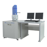
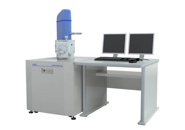
【DISCONTINUED】JSM-6510 Series Scanning Electron Microscope
DISCONTINUED
This product is no longer available.
If you would like to know the latest information about your preferred product or to find out more about alternatives, please click on the link below. We hope you will continue to use our products.
A general-purpose, thermal type SEM to meet the needs of a wide range of users with built-in standard recipes. A wealth of available options, such as Cryo, further expands the range of applications.
Features
Operation navigation
The operation navigation screen displays a variety of navigation functions.
The specimen exchange procedure is presented in a flow format, so that the process can be easily completed even by a first time user.
When the motor-drive stage option installed, a stage graphic makes it easy to search for a target site When the stage navigation system (option) is used, it is possible to locate a region of interest with the feel of operating an optical microscope.
Video animations supplement the maintenance procedure explanations
Standard recipes
Standard recipes incorporating JEOL expertise and experience are pre-installed
Even new SEM users can set conditions suitable for each sample
Wide range of options
A variety of options can be installed, including a cryo system and a cool stage
Since the EDS is also a JEOL product, control can be performed from a single PC
EDS integration
Analysis can be started from the SEM window
Since the EDS is also a JEOL product, control can be performed from a single PC
Specifications
| ResolutionHV mode | 3.0 nm(30 kV)、8 nm(3 kV)、15 nm(1 kV) |
|---|---|
| LV mode *1 | 4.0 nm(30 kV) |
| Magnification | × 5 to × 300,000 (on 128 mm × 96 mm image size) |
| Preset magnifications | 5 step, user selectable |
| Standard recipe | Built in |
| Custom recipe | Operation conditions (Optics, Image mode, LV pressure*1) Specimen stage |
| Image mode | Secondary electron image, REF image, Composition*1, Topography*1, Shadowed*1 |
| Accelerating voltage | 0.5 kV to 30 kV |
| Filament | Factory pre-centered filament |
| Electron gun | Fully automated, manual override |
| Condenser lens | Zoom condenser lens |
| Objective lens | Super conical objective lens |
| Objective lens apertures | 3 stages, XY fine adjustable |
| Stigmator memory | Built in |
| Electrical image shift | ± 50 μm (WD = 10 mm) |
| Auto functions | Focus, brightness, contrast, stigmator |
| Specimen stage | Eucentric large-specimen stage
X: 80 mm, Y: 40 mm, Z: 5 mm to 48 mm, Tilt: −10° to 90°, Rotation: 360° |
| Reference image (Navigator*3) | 4 images |
| Specimen exchange | Draw out the stage |
| Maximum specimen | 150 mm diameter |
| PC | IBM PC/AT compatible |
| OS | Windows 7 |
| Monitor | 19 inch LCD, 1 or 2*2 |
| Frame store | 640 × 480, 1,280 × 960, 2,560 × 1,920, 5,120 × 3,340 |
| Dual live image | Built in |
| Full size image display | Built in |
| Pseudo color | Built in |
| Multi image display | 2 images, 4 images |
| Digital zoom | Built in |
| Dual magniἀcation | Built in |
| Network | Ethernet |
| Measurement | Built in |
| Image format | BMP、TIFF、JPEG |
| Auto image archiving | Built in |
| Pumping system | Fully automated, DP: 1, RP: 1 or 2*1 |
| Switching vacuum mode*1 | Through the menu, less than 1 minute |
| LV Pressure*1 | 10 to 270 Pa |
| JED-2300 EDS*2 | Built in |
Principal Options
Backscattered electron detector*1
Low vacuum secondary electron detector
Energy dispersive X-ray analyzer (EDS)
Wave length dispersive X-ray analyzer (WDS)
EBSD
Stage navigation system
Airlock chamber
Chamber scope
Operation keyboard
LaB6 electron gun
Report creation software (SMile View™)*2
Operation console (750 mm wide, 900 mm wide)
Motor controlled stage (2 axes, 3 axes, 5 axes)
Standard on JSM-6510LA and JSM-6510LV
Standard on JSM-6510LA and JSM-6510A
Available when the motorized specimen stage is provided.
Application
Application JSM-6510series
High Angle Backscattered Electrons and Low Angle Backscattered Electrons
Introducing the ALTO Series (Cryo System)
Introducing Cryo Scanning Electron Microscopy
Gallery
More Info
Are you a medical professional or personnel engaged in medical care?
No
Please be reminded that these pages are not intended to provide the general public with information about the products.