Release of the New Scanning Electron Microscope JSM-IT510 Series InTouchScope(TM) - Easy to acquire data for all specimen types -
Release Date: 2021/11/08
JEOL Ltd. (President & COO Izumi Oi) announces the development and release of a new Scanning Electron Microscope (SEM), the JSM-IT510 series, in November 2021.
Product Development Background
Scanning electron microscopes are used in a wide range of fields, such as nanotechnology, metals, semiconductors, ceramics, medicine, and biology. In addition, SEM applications are expanding to not only basic research, but also quality control at manufacturing sites. With this, demands for faster and easier data acquisition of both SEM images and analysis results, such as energy dispersive X-ray spectroscopy (EDS) spectra, are increasing.
In order to meet these needs and increase throughput, we have developed the JSM-IT510 series, which further evolves the operability of our popular InTouchScope™. With the new Simple SEM function, you can now "leave" your daily routine (repetitive operation) to the instrument.
Main Features
- New "Simple SEM" function
The Simple SEM function allows the user to simply select the acquisition conditions and field of view for the SEM image, and then the SEM image is automatically acquired. Routine work can be made more efficient. - New "Low-vacuum Hybrid Secondary Electron Detector (LHSED)"
This new detector collects both electron and photon signals providing an image with high S/N and enhanced topographic information even under a low vacuum. - Integration of the Scanning Electron Microscope (SEM) and Energy Dispersive X-ray Spectrometer (EDS) System
The integration of SEM and EDS has been further developed, and the Live Map function enables live display of the elemental map of the observation field of view. - New "Live 3D" function
3D images can be constructed on the spot while SEM observation is being performed to obtain unevenness and depth information. - Live Analysis function
The embedded EDS system shows a real-time EDS spectrum during image observation for efficient elemental analysis. - New Stage Navigation System LS function
The new Stage Navigation System LS can acquire an optical image of an area four times larger than that of conventional models (200 mm x 200 mm). This function allows the user to acquire an optical image of the observation sample and move to the desired observation field by simply clicking on the optical image. - Zeromag
With our Zeromag function, sample navigation is even easier than ever. You can locate areas for imaging or specify analysis positions over multiple fields using an optical image or holder graphic. - Display of the characteristic X-ray generation depth
This supports a prompt understanding of the analysis depth (reference) for the specimen. - SMILE VIEW™ Lab, enabling integrated management of image and analysis data
Facilitates report generation for all data from collected SEM images to elemental analysis results in a short time.
Main Specifications
| Resolution HV mode | 3.0 nm (30 kV) 15.0 nm (1.0 kV) |
| Resolution LV mode | 4.0 nm (30 kV with Backscattered electron image) |
| Direct magnification | ×5 to ×300,000 (Defined with a display size of 128 mm×96 mm) |
| Displayed magnification | ×14 to ×839,724 (Defined with a display size of 358 mm×269 mm) |
| Electron gun | Tungsten (W) filament |
| Accelerating voltage | 0.3 kV to 30 kV |
| Probe current | 1 pA to 1 μA |
| LV pressure adjustment | 10 to 650 Pa |
| Automatic functions | Filament adjustment, Gun alignment, Focus / Stigmator / Brightness / Contrast |
| Maximum specimen size | 200 mm diameter, 90 mm height |
| Specimen stage | Large eucentric type X: 125 mm Y: 100 mm Z: 80 mm Tilt: -10 to 90° Rotation: 360° |
| Standard recipe | Built-in (includes EDS conditions) |
| Image mode | Secondary electron image, REF image, Compositional image, Topographic image, Stereo-microscopic image |
| EDS functions | Spectral analysis, Qualitative & Quantitative analysis, Line analysis (horizontal line, specific direction line), Elemental mapping, Probe tracking |
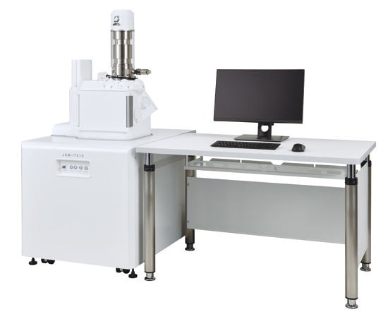
Note: Specification and a photo of the instrument are those of JSM-IT510 (LA)
Sales target
200 units / year
Link
Are you a medical professional or personnel engaged in medical care?
No
Please be reminded that these pages are not intended to provide the general public with information about the products.