Volume EM ~ 3D Reconstruction ~
This is a technique of observation and analysis of the inner structure of a specimen using 3D reconstruction by using electron microscopes. There are two 3D observation methods using electron microscopes: the first method is the 3D reconstruction method that stacks successionally captured cross-sectional images (array tomography, serial block face, FIB-SEM)., and another method is 3D reconstruction by calculations from projection images of the specimen at continuously tilts (TEM tomography).
For details of each technique, click here to see.
Array Tomography
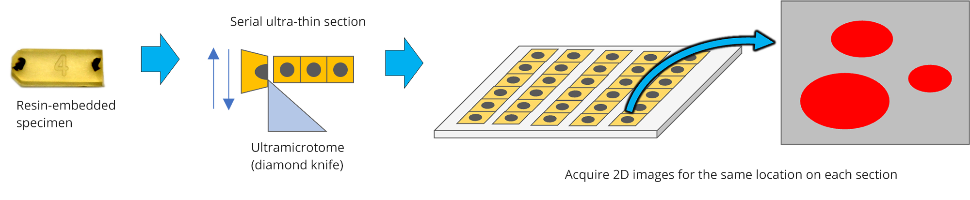
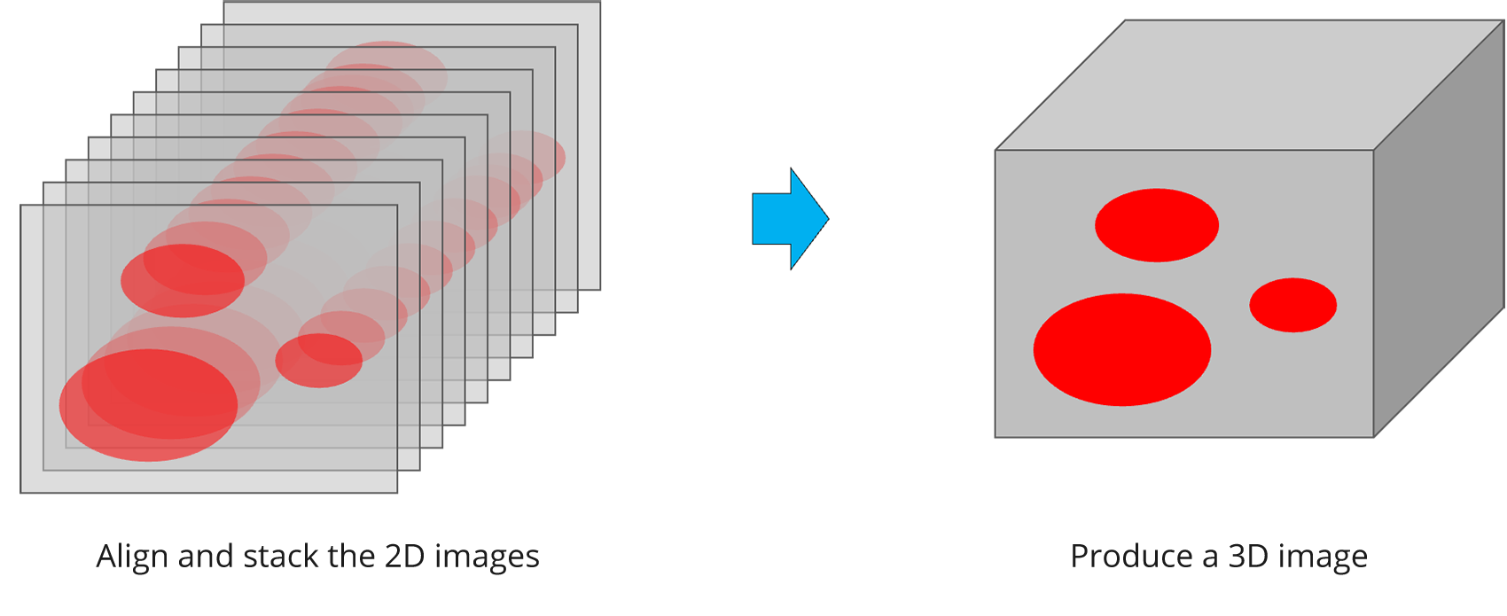
Array tomography provides three-dimensional information of specimens such as biological tissues. The specimen embedded in resin is sliced into serial sections. The same positions of the sections are observed using scanning electron microscope (SEM) to make a stack of images, which is used for three dimensional reconstruction.
Array Tomography Workflow
Serial Block Face (SBF)
The surface of the biological specimen that is embedded in resin is cut by a diamond knife and observed by a
SEM. By repeating the cutting and observation, the SEM image series can be obtained in the thickness
direction. A three-dimensional reconstruction is performed by stacking this SEM image series.. Further,
extraction(segmentation) of the target object (entire cells, organelles, etc) reveals a 3D structure in the
hundreds μms range.
Cutting and observations can be performed automatically with the specialized slicing device that is built in
the SEM specimen chamber.
* Deerinck, T. J., Bushong, E., Thor, A. & Ellisman, M.H. NCMIR methods for 3D EM: A new protocol for preparation of biological specimens for serial block-face SEM. Microscopy (Oxf),6-8. (2010)
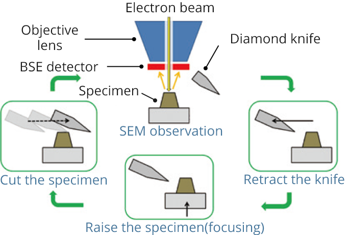
Workflow of cutting & observation
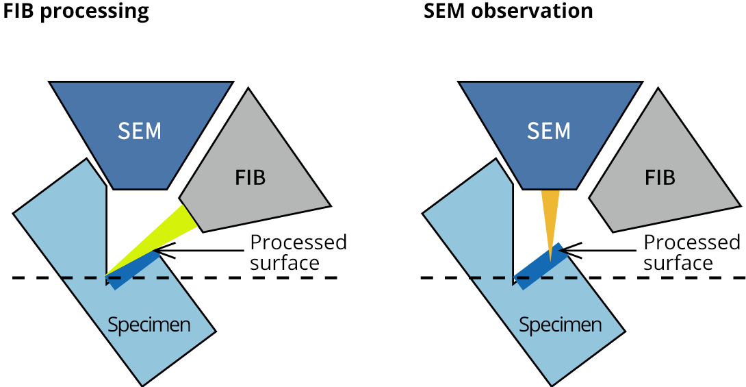
FIB-SEM Method 3D Reconstruction
Cross sectioning with an FIB and obtaining an SEM image are performed serially. A 3D reconstruction is performed by stacking the serial cross-sectional SEM images. This method requires samples to be stained with powerful heavy metals and embedded in resin. The advantages of this method are high positional accuracy, small distortion due to processing, high cutting resolution (less than 20 nm), and the ability to cut hard materials such as teeth, bones, and metal implants that are difficult to cut with a diamond knife.
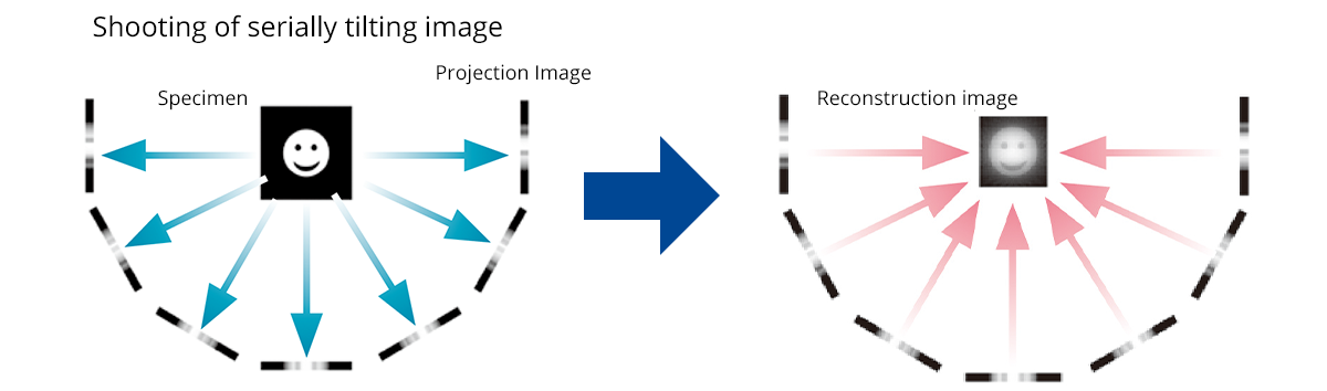
TEM Tomography
The thickness of the samples observed with a TEM is 100 nm or less, but inside them there are fine structures smaller than the thickness of the sample. 3D reconstruction with the TEM Tomography Method can reconstruct 3D structures inside the specimen with high resolution by calculation using projection images taken at continuous tilts of the sample.
Click the button below to return to the TOP page of Biology / Life Science