CLEM for Life Science
What is CLEM
CLEM(Correlative Light and Electron Microscopy)is a method for effective observation and analysis by integrating information of the same specimen obtained by a light microscope(LM) and transmission electron microscope(TEM) or scanning electron microscope(SEM). The advantages of both can be utilized by doing this for observation and analysis.
Please click here and check each method of CLEM for Life Science.
polish CLEM / milling CLEM
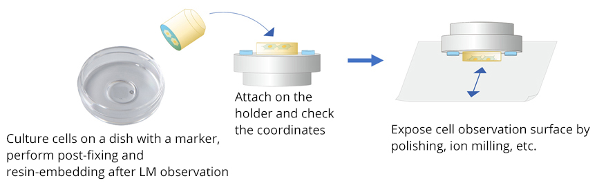
This is a method to observe the same field of view as an LM image (optical image) by using a scanning electron microscope by exposing it to the surface by polishing or ion milling, etc. It is possible to observe the entire image or fluorescent staining image of cells by using LM, and then observe the detailed structure inside the cell using SEM. The capability of simple and extensive observation is the feature. The use of a specialized linkage holder makes it easy to align the position.
Polish CLEM / milling CLEM workflow
Cells are cultured on a dish with a marker and pre-fixed and stained . After an LM observation, the preparation sample is attached to the holder and the coordinates are confirmed. The observation surface is exposed by polishing and ion milling, and observed by using an SEM.
Application examples for which this technique is suitable
- Observation of structure inside a cell
- Wide field of view observation
- Multi-point observation
on chip CLEM
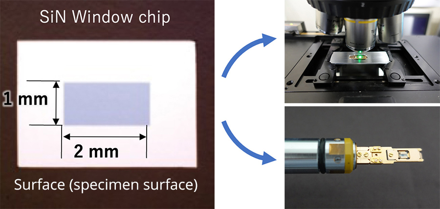
This is a method to observe an ultra-thin specimen on the same specimen mount (SiN window chip) by using both a light microscope (LM) and transmission electron microscope (TEM). Because there is no process to cause a deformation of a specimen between observations by LM and TEM, highly accurate overlaying is possible. This method can be also used for the material field.
on chip CLEM workflow
The on chip CLEM is to put the ultra-thin section specimen prepared for TEM on a SiN
window chip. The SiN window chip can be attached to the holders for the light microscope, and TEM by using a
specialized retainer. By switching the use of a specialized retainer to the LM holder and TEM holder, LM・TEM
observation can be easily performed.
Since the same sections can be observed, highly accurate
correlations can be obtained.
Application examples for which this technique is suitable
- Specimen in which fluorescence or special scattered light can be observed after an ultra-thin section preparation
- Specimen in which phase difference observation can be performed
- Toner particles (with autofluorescence), or a crystalline polymer,
etc.
other than a biological specimen
LLP-CLEM
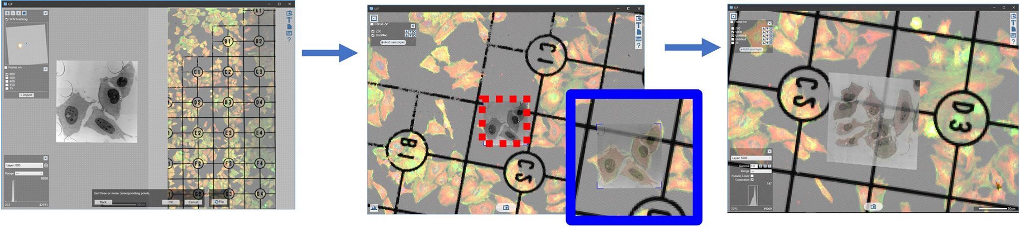
Limitless Panorama (LLP) has a function to import images taken externally, such as with a light microscope. LLP can match the area of observation of LLP with the specimen coordinates of a transmission electron microscope. After matching the coordinates of each observation image, a montage pictures of the area that was observed by LM, are taken by using the TEM. A wide range can be observed with high resolution.
LLP-CLEM Workflow
Cells cultured on a dish with markers are pre-fixed and stained. After the LM
imaging, the target area of the specimen after post-fixation and resin-embedding is cut out using a marker
as a landmark. An ultra-thin section of the specimen is prepared by using ultramicrotome and observed by
using the TEM.
Alignment is performed by specifying three or more feature points in common between the LM
image that was taken and the TEM image of the low magnification image. Specify an area and perform LLP
imaging.
Application examples for which this technique is suitable
- Wide field of view observation
- Searching for small amount of mutant cell, etc.
In resin CLEM
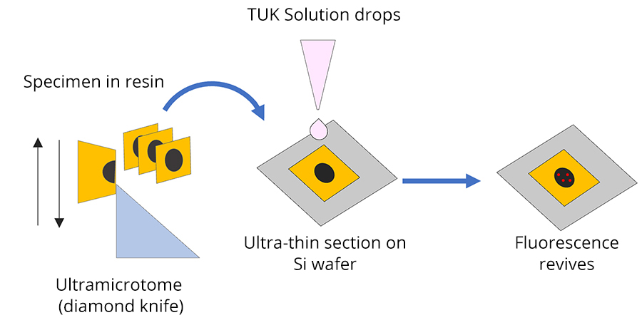
In resin CLEM is a technique to prepare an ultra-thin section after fluorescent staining and resin-embedding of the specimen, revive the fluorescence, and observe the same section by using a light microscope and electron microscopes (TEM/SEM). To revive the fluorescence, a fluorescence reviver (TUK solution) is used. The same section can be observed with both light and electron microscopes, allowing highly accurate superposition of optical and electron microscopic images.
In resin CLEM workflow
An ultra-thin section is mounted on the SiN window chip (for TEM) or Si wafer or slide glass (for SEM) that has a smooth surface. Fluorescent reviver (TUK solution) is applied and observed by a light microscope, followed by an electron microscope.
Application examples for which this technique is suitable
- Fluorescent protein whose fluorescence can revive with the use of fluorescence reviver and the cultured cells and tissues where the introduction of fluorescent dyes is possible.
- Sample that requires highly accurate alignment such as localization of proteins in an organelle.
Multi-color CLEM
Various modifications are possible with organic silica nano particles, such as mixing fluorescent dyes or adding heavy metals. Organic silica nano particles with fluorescent dyes and heavy metal are taken into cells. The organic silica nano particles with several kinds of heavy metals can be distinguished by the heavy metal added through EDS mapping. Thus, it is possible to observe it in multiple colors not only by light microscopes, but also by electron microscopes. It is also possible to use this as a marking when the area observed by a fluorescent microscope is observed by an electron microscope in detail.
Multi-color CLEM workflow
Organic silica nano particles are modified such as "green fluorescence + gold particle", and "red fluorescence + iron particle". This is introduced to the cultured cell and then confirmation of the same area is possible by using fluorescence observation and EDS mapping.
Application examples for which this technique is suitable
- Macrophage-type cells phagocytosing silica nanoparticles
- Microorganisms eating silica nano particles such as paramecium and amoeba
- Marking is possible for cells in vivo through intravenous injections
Introduction of Related Products
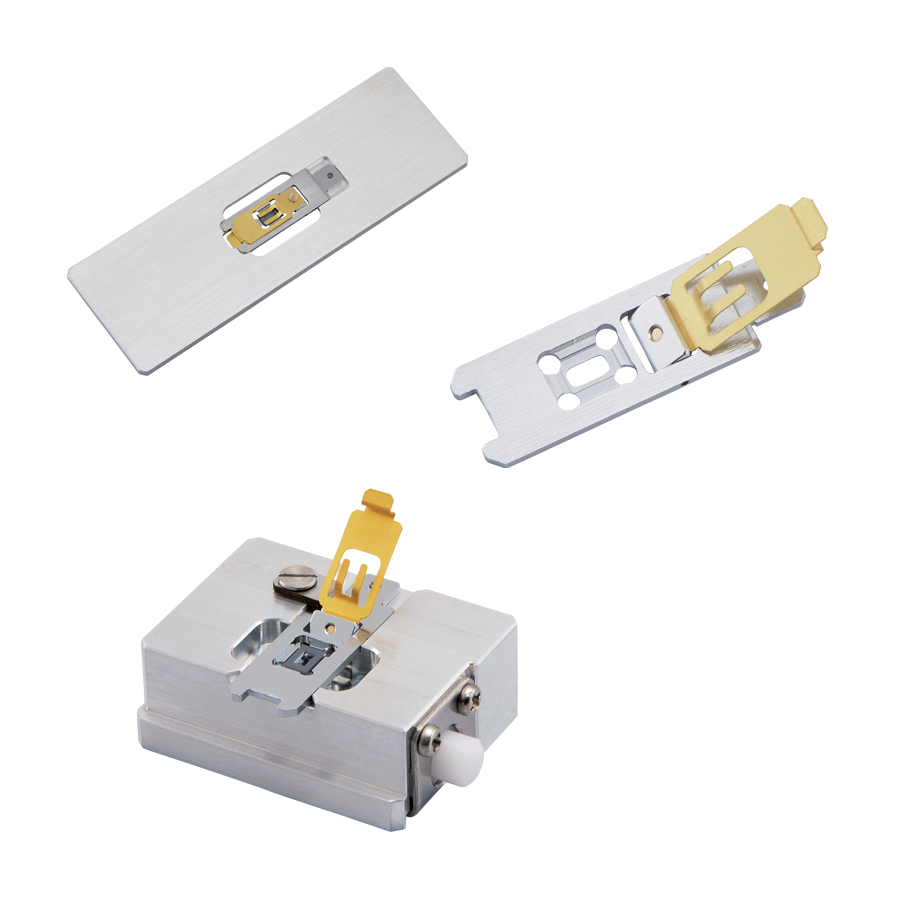
SiN Window Chip
The high-strength SiN film enables us to observe a serially large area of a millimeter in size. It is also ideal for observation of serially sliced sections because there is no invisible area that is caused by conventional TEM grids. The dedicated retainer makes it easy to perform Correlative Light and Electron Microscopy (CLEM).
Click the button below to return to the TOP page of Biology / Life Science