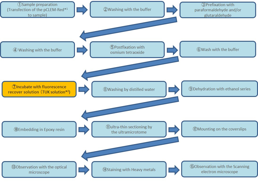
In resin CLEM with fluorescence recover solution
SM2021-03E
Recently, various physiological phenomena and localizations of biological substances have been observed with optical microscopes by using fluorescent dyes and fluorescent proteins. CLEM is a method of observing the same sample with optical microscopes and electron microscopes, and merging these observation images to integrate information obtained by each instrument. Therefore, high accuracy alignment is required for this image merging. However, the fluorescence of fluorescent dyes and proteins observed with an optical microscope is quenching by sample preparation for electron microscope observation (fixation, dehydration, resin embedding, etc), Optical microscope observation and electron microscope observation had to be performed under different conditions. CLEM-Red*1 is a fluorescent protein that recovers fluorescence by treatment with a fluorescence recover solution (TUK solution*2) even after fixation with glutaraldehyde or osmium tetroxide, and this fluorescence keeps even after embedding in resin. By using CLEM-Red and the fluorescence recover solution, we can observe ultra-thin sections after resin embedding with fluorescence microscopes, which enables highly accurate CLEM.
Protocol
*1,2 CLEM-Red, TUK solution are products of Fujifilm Wako Pure Chemical Corporation.
Results
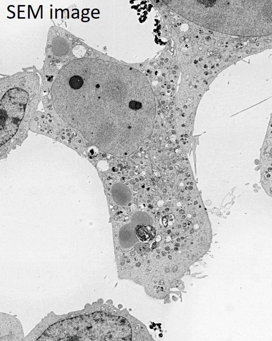
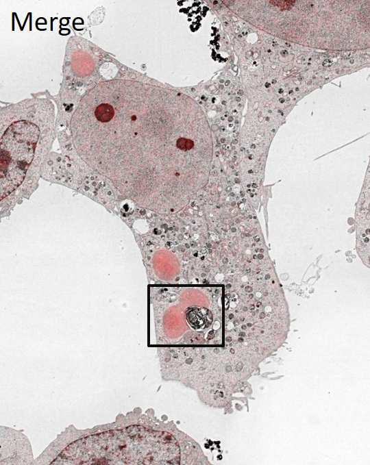
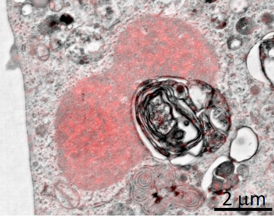
The phenomenon of degradation for unnecessary organelles and proteins in cells is called autophagy. In this autophagy, the autophagosome that is the membrane structure surrounds unnecessary substances is observed. It has been suggested that the p62 protein plays an important role in the formation of this autophagosome. HeLa cells overexpressing the fusion protein of p62 and CLEM-Red were observed in Resin CLEM.
As a result of this observation, the CLEM-red-p62 fusion protein was localized in the oil droplet structure of the cell, and autophagosomes were formed in the adjacent region.
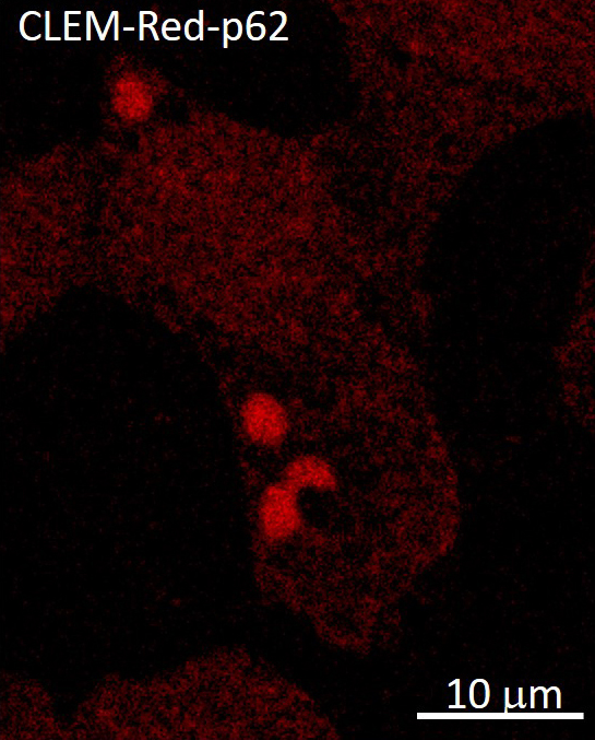
Sample: Hela Cell, pCLEM-Red-p62
Confocal microscope: A1 (Nikon)
Scanning electron microscope: JSM-7900F (JEOL)
Sample provision
Dr. Isei Tanida, Prof. Yasuo Uchiyama (Juntendo university, Tokyo)
Reference
Tanida I, Haruta T, Suga M, Takei S, Takebe A, Furuta Y, Yamaguchi J, Oliva
Trejo JA, Kakuta S, Uchiyama Y. Membranous Structures Directly Come in Contact
With p62/SQSTM1 Bodies.
J Histochem Cytochem. 2021 Jun; 69 (6) : 407-414.
- Please see the PDF file for the additional information.
Another window opens when you click. 
PDF 1.3 MB
Are you a medical professional or personnel engaged in medical care?
No
Please be reminded that these pages are not intended to provide the general public with information about the products.