Radiation Dose Measurement by ESR of Teeth
ER090003E
Bones and teeth consist of microcrystalline hydroxyapatite Ca10(PO4)6(OH)2, and organic protein,collagen. The enamel covering the surface of teeth consists mainly of hydroxyapatite and is very resistant to metabolic change, therefore can be used as a very sensitive dosimeter in measuring radiation exposure.
In general, the radiation dose is obtained using the signal intensity of the CO2- radical generated by irradiation of the carbonate ion CO32-, which is contained in the enamel as an impurity. It is claimed that measurement as low as 30 mGy is possible (1).
However, for doses less than 5 Gy, as the signal of organic radicals (g=2.0046) is superimposed on the signal of the CO2- radical, this contribution must be subtracted (2) (3). If a sample is heated, the ESR signal of the organic radical which is generated by the decomposition of collagen protein increases (4). The reason why only enamel is generally used as the sample for the ESR method is because the g-value of the organic radical is almost the same as that of the CO2- radical. Therefore, when the evaluation of low dose samples is calculated, it is necessary to separate spectra and subtract the signal of the organic radical, otherwise it could lead to an overestimate. Also, it is necessary to check if there is a contribution to radiation exposure by dental x-rays. There are many studies on the ESR Dose Measurement Using Enamel of Teeth at several institutes (5),(6),(7). Please refer to “Application Note ER-080001” on how to calculate the exposed dose.
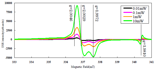
Fig.1. Change of Tooth’ ESR Signal by Microwave Power
(Using 60Co radiation source of JAERI Takasaki Laboratory, and irradiate g rays for about 2.5kGy)
Sample courtesy of Dr. Shitaoka of Nara University of Education
References
- Ikeya M. (1987): ESR(Electron Spin Resonance)Dating. IONICS , p210.
Ikeya M. (1993): “New Applications of Electron Spin Resonance. Dating, Dosimetry and Microscopy.” World Scientific , p.500. - (1) Romanyukha A. A. et al (2005) : Parameters affecting EPR dose reconstruction in teeth. Applied Radiation and Isotopes, Vol.62, 147-154.
- (2) Ivannikov A. et al (2000) : EPR tooth enamel dosimetry: Optimization of the automated spectra deconvolution routine. Health Physics, 81, 1245-137.
- (3) Toyoda S. et al (2003) : Gamma-ray dose response of ESR signals in tooth enamel of cows and mice in comparison with human teeth. Radiation Measurements, 37, 341-346.
- (4) Caddie D. A . et al (1985) : ESR considerations in the dating of Holocene and late Pleistocene bone material. ESR Dating and Dosimetry, 353-361.
- (5) Chumaka V. et al (1996) : The first international intercomparison of EPR-dosimetry with teeth: First results. Applied Radiation and Isotopes, 47, 1281-1286.
- (6) Wieser A. et al (2000) : The second international intercomparison on EPR tooth dosimetry. Radiation Measurements, 32, 549-557.
- (7) Wieser. A et al (2005) : The 3rd international intercomparison on EPR tooth dosimetry: Part 1, general analysis. Applied Radiation and Isotopes, 62, 163-171.
SEARCH APPLICATIONS
Related Products
Are you a medical professional or personnel engaged in medical care?
No
Please be reminded that these pages are not intended to provide the general public with information about the products.