3-dimensional Structure with high precision TEM Tomograph System is an automation system handling an entire process from an acquisition of sequential tilt images through to their 3-dimensional reconstruction. Its software (Recorder, Composer, Visualizer-Kai) employ unique algorithms for enabling the automation of various adjustments particularly required to Tomography.
Features
Recorder
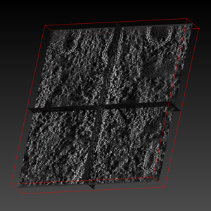
This software acquires sequential tilt images in a short time for Tomography. Many images of samples, taken in the course of continuous tilting with certain angle step, are necessary for accurate reconstruction. Operations such as sample tilting at a pre-set angle step, correction of view shift stemming from the tilt, adjustment of focusing and the acquisition of images are automatically carried out.
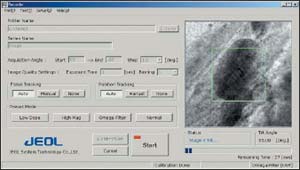
Recorder Operation Screen
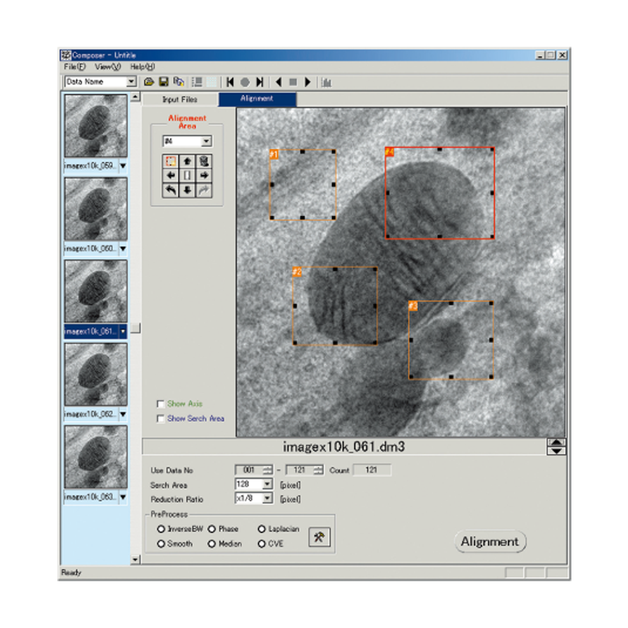
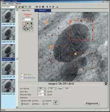
Composer
This software performs a 3-dimensional reconstruction from sequentially acquired tilt images. Crucial element required to the electron beam tomography is how to make an accurate alignment of the image data. This is resolved by calculation of 3-dimensional position on the tilt axis and analyzing the precise angle information, and as a result, a precise reconstruction in a short time with easy operation has been realized.
Visualizer-Kai
This software displays 3-dimensionally-reconstructed data by various methods such as volume rendering and surface rendering and also enables the intuitive observation on the screen as if handling by hands. By using a mouse, the images can be freely moved and displayed. The function of length measurement is also included.
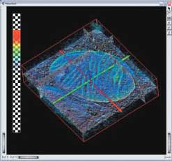
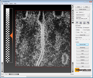
Visualizer-Kai Operation Screen
High Tilt Sample Stage (optional)
To acquire image at high tilt angle, high tilt sample stage is needed.
Application Example of 3-dimensional Reconstruction: 3-dimensional structure analysis of a Tight junction
Measurement Condition
Accelerating Voltage: 120kV
Tilt angle: ±65°, 1° step
Sample: Tight junction (mouse’s small intestine epithelium, thickness 70nm)
Explanation: Tight junction is a cell adhesion structure existing in the top area of epithelium cell of vertebrata.
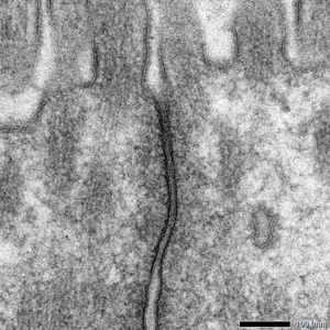
Fig. 1 TEM Image of Tight Junction
Tight junction works as a barrier against water or ions, as its cell membranes between neighboring cells closely adhere. This time, we analyzed a 3-dimensional form of a cell membrane at the Tight junction.
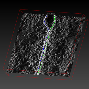
Movie 1: Positioning of image data by Composer
By using Composer, we adjusted the position of sequential tilt image photographed by Recorder. Although a fiducial marker such as an Au colloid particle was needed in conventional method, Composer realizes positioning without it.
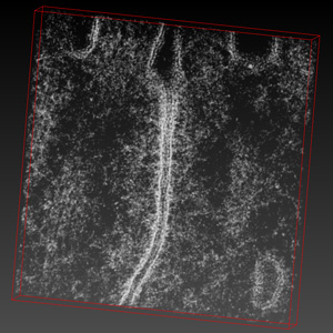
Volume rendering image of tight junction
Movie2: volume-rendering image of tight junction
By using Visualizer-Kai the 3-dimensionally reconstructed image can be freely moved and observed.
Slice image of tight junction
Movie 3: slice image of tight junction
We can observe that the adhered structure of the cell membranes at a tight junction is a dynamic structure where adhering position changes 3-dimensionally, rather than the structure where membranes at the same position keep adhering.
Length measurement
Visualizer-Kai has length-measurement function.
Application
Structural Analysis of Semiconductor Devices by Using STEM/EDS Tomography
More Info
Are you a medical professional or personnel engaged in medical care?
No
Please be reminded that these pages are not intended to provide the general public with information about the products.