SM-92100EUVC
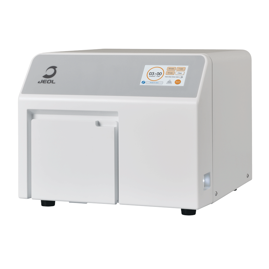
EXCIMER UV CLEANER
Features
Mitigates Contamination during Observations Made with an Electron Microscope
Cleans Specimens and Tools
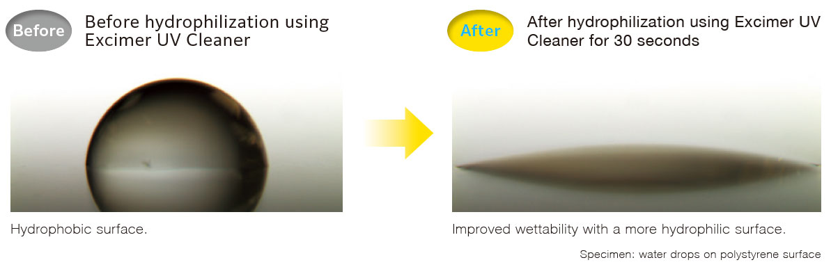
Hydrophilizes the Specimen Surface
Dark areas of contamination can gradually form on a specimen surface during observation in an electron microscope.
This surface contamination can conceal the true surface structure of the specimen or degrade resolution in the contaminated area.
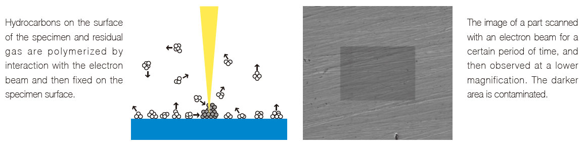
Ever see that 'Dark Square'? This happens when hydrocarbon-based contaminants on the surface of the specimen are irradiated by the electron beam resulting in carbonaceous deposits.
The Excimer UV Cleaner uses UV light (Xe excitation, at 172 nm) to eliminate contamination on the specimen surface. This system can also be used to hydrophilize a specimen surface.
Mitigate Contamination during Observation
Observation of fine surface structure in SEM requires imaging at low accelerating voltage. Under these conditions,
surface contamination can conceal the observation of this fine structure.
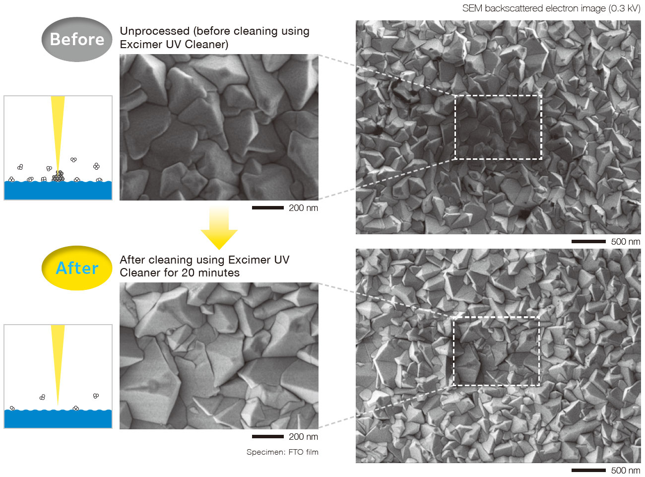
Observation was performed at a magnification of 100,000 times (left figure), then at a lower magnification of 30,000 times (right figure). When the specimen is not processed (top row), the part observed at 100,000 times turns darker and less clear. However, after it is cleaned using the Excimer UV Cleaner (bottom row), contamination caused by irradiation of an electron beam is less, because there are fewer hydrocarbon compounds on the specimen surface.
Cleaning Contamination Adhered to the Specimen Surface
If a specimen is used or stored for a long time, contamination accumulates due to organic compounds contained in the environment.
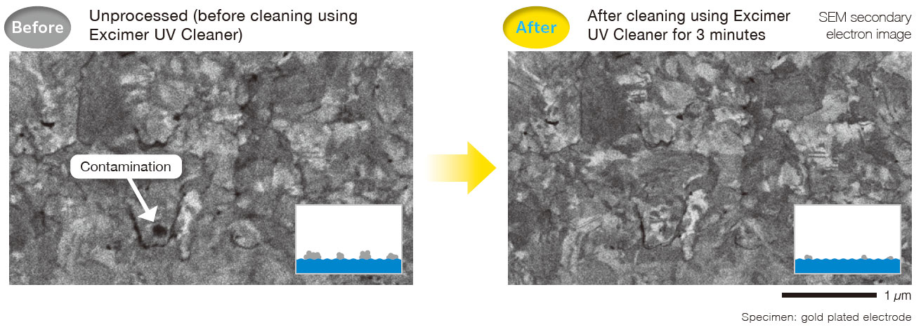
The Excimer UV Cleaner can eliminate contamination caused by organic substances in a dry environment.
Hydrophilization
When the Excimer UV Cleaner irradiates UV light, reactive oxygen species are generated and can form polar functional groups on the surface
which improves hydrophilicity. This pre-treatment process can be applied to improve bonding when processing a specimen for EM observation.
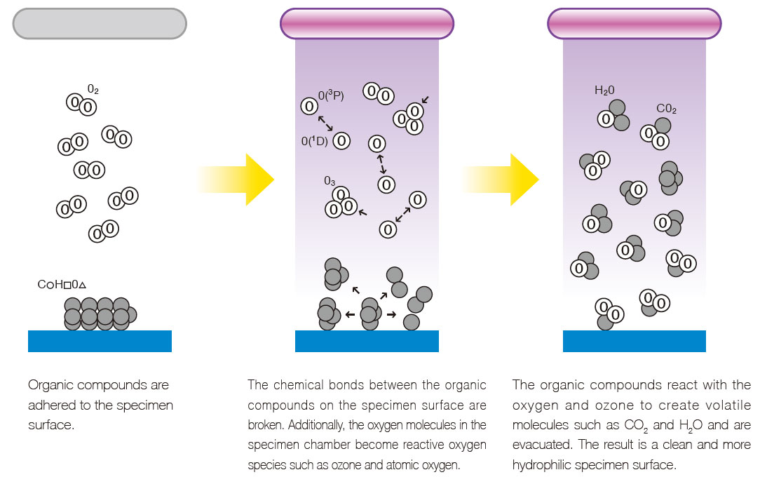
Mechanism for Specimen Cleaning
/ Hydrophilization Using the Excimer UV Cleaner
Process of contamination caused by the electron beam
Cleaning process
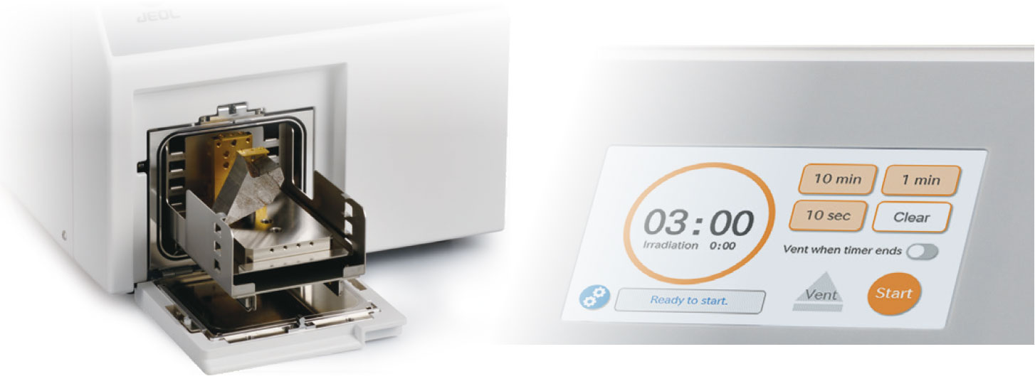
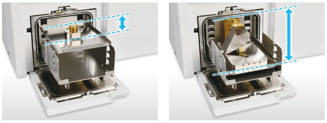
The height of the specimen tray can be adjusted to fit large specimens.
Specifications
| Wavelength of irradiated UV light | 172 nm | ||
|---|---|---|---|
| Maximum specimen size | 105 mm (W) × 105 mm (D) × 70 mm (H) | ||
| Maximum specimen mass | 1 kg | ||
| Irradiation time | 10 seconds to 99 minutes | ||
| Vacuum pump | Diaphragm type dry vacuum pump | ||
| Installation requirements: |
Power | Single phase 90 to 110 V AC, 50 / 60 Hz, 500 VA | |
| Dimensions and weight (cables and vacuum pipes not included) |
Main unit | 39 cm (W) × 50 cm (D) × 32 cm (H), approximately 22 kg | |
| Vacuum pump | 50 cm (W) × 20 cm (D) × 30 cm (H), approximately 9 kg | ||
| Room temperature | 15 to 30 ℃ | ||
| Humidity | 60% (RH) or less (no condensation) | ||
| Regular replacement parts (replace them every time cumulative irradiation time reaches 1000 hours) | UV lamp, vacuum gauge, O-ring, etc. | ||
* Note: Specifications are guaranteed when no modification or addition is made and are subject to change without notice.
Catalogue Download
SM-92100EUVC EXCIMER UV CLEANER
Related Products
Are you a medical professional or personnel engaged in medical care?
No
Please be reminded that these pages are not intended to provide the general public with information about the products.