


Introduction to Multinuclear NMR│NMR Measuring Nuclei Beyond 1H and 13C
Multinuclear NMR is a powerful analytical technique that enables the observation of a wide variety of nuclei such as15N, 11B, 19F, and 31P. However, because each nucleus has distinct sensitivity, relaxation properties, and measurement requirements, an understanding of proper instrument setup and experimental conditions is essential for obtaining reliable results. In this column, we will explain the key points necessary for successful multinuclear NMR experiments such as the fundamentals of multinuclear NMR, the factors that influence detection sensitivity, and pulse conditions in a clear and accessible manner.
What is multinuclear NMR?
Multinuclear NMR is a generic term for NMR measurements of nuclei other than the commonly measured 1H and 13C. Since modern NMR spectrometers are digitally controlled, in many cases, the observed nucleus can be changed simply the software settings. A wide variety of nuclei can be measured although the measurable nuclei depend on the probe (detector) and the type of spectrometer.
Why is multinuclear NMR required?
NMR is widely used as a routine analytical tool in organic chemistry, particularly through 1H NMR
and 13C NMR. However, in multidisciplinary fields such as coordination chemistry, biochemistry,
and materials science, there are many situations in which information about nuclei other than the commonly
observed nuclei, such as hydrogen and carbon is required. These include elements such as phosphorus,
nitrogen, fluorine, silicon, and lithium that are present within molecules.
By using multinuclear NMR, it becomes possible to obtain information such as the following.
- Coordination states of phosphate esters and organophosphorus compounds (31P)
- Chemical environment of nitrogen in amide group of protein and biomolecule (15N)
- Structure confirmation of fluorine-containing pharmaceuticals and metabolic tracking (19F)
- Si bonding environments in inorganic materials such as silicones and zeolites (29Si)
- Local structure of lithium ion battery electrolyte (7Li)
POINT
Multinuclear NMR is not a special technique but rather an application of the same principles used in 1H NMR to other nuclei. However, sensitivity, measurement condition, and the required instrument configuration greatly differ for each nucleus. The key to successful analysis is to understand the "characteristic" of each nucleus.
Review of NMR-detectable and non-detectable nuclei
Spin quantum number and NMR observability
To observe NMR signals, the target nucleus must have a nonzero nuclear spin. This property is expressed by the spin quantum number I, and I ≠ 0 is a necessary condition for NMR observation.
Spin quantum number is determined by the combination of the number of protons and the number of neutrons, and follows the rule shown below:
| Number of protons | Number of neutrons | Spin quantum number I | Observability | Example |
|---|---|---|---|---|
| Even numbers | Even numbers | I = 0 | Not observable | 12C, 16O, 32S |
| Odd numbers | Even numbers / Odd numbers | I = 1/2 | Observable | 1H, 13C, 19F, 31P |
| Odd numbers or Even numbers | Odd numbers or Even numbers | I > 1/2 (half-integer) | Depending on the conditions | 11B, 23Na, 35Cl |
| Odd numbers | Odd numbers | I = Integer ( ≥ 1) | Depending on the conditions | 2H, 14N |
Why nuclei (I = 0) with even numbers of protons and neutrons cannot be observed by NMR
For nuclei in which both the proton number and neutron number are even (such as 12C and 16O), the nucleons form pairs and cancel each other's spins. As a result, the total nuclear spin becomes zero (I = 0). Since NMR utilizes the phenomenon in which the energy levels of nuclear spins split in a magnetic field, this splitting does not occur in the nuclei with zero nuclear spin, and therefore resonance with radiofrequency radiation cannot be generated.
Why NMR observation of paramagnetic species is difficult
When there are paramagnetic ions with unpaired electrons (Fe3+, Cu2+, Gd3+,
etc.) present in the sample, the large magnetic moment of the electron spin strongly affects the surrounding
nuclei. As a result, relaxation of the nuclear spin becomes extremely fast. As relaxation becomes fast, the
NMR signal (FID) decreases in a very short time. Consequently, the peaks become broadened (broadening), and
the NMR signal becomes difficult to observe depending on the situation.
In addition, even if the NMR signal is observed, it may appear far from its normal chemical shift position
(paramagnetic shift). Thus, the NMR measurement of the paramagnetic atomic species is extremely difficult to
handle.
Fundamental physical properties of target nuclei to check before multinuclear NMR
How to read the "NMR periodic table"
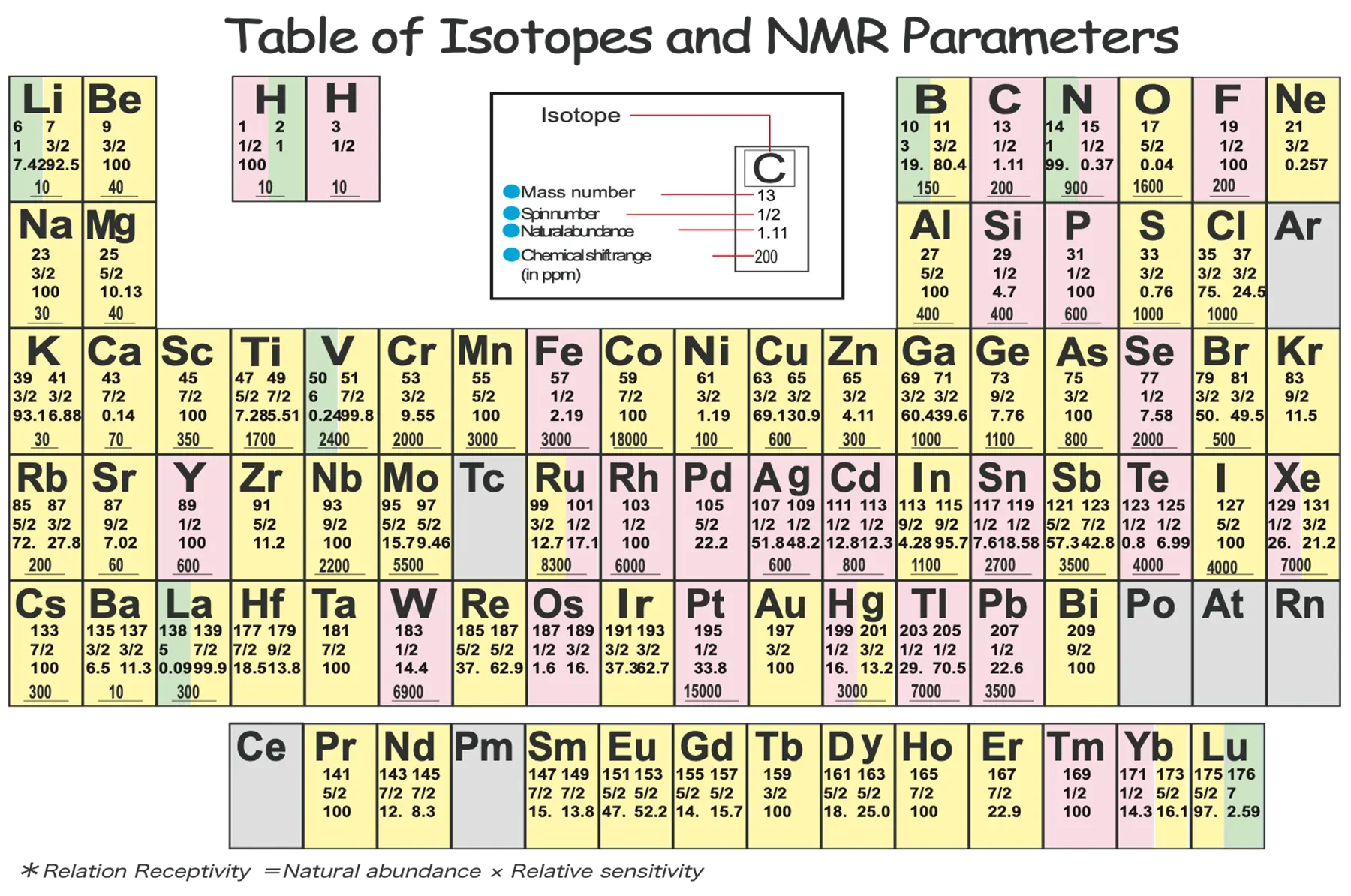
When starting multinuclear NMR measurements, an NMR periodic table is a useful reference which
differentiates the spin quantum number and the natural abundance by color.
For example, the carbon is shown with the mass number 13 on the periodic table above. Generally, the most
abundant isotope of carbon is 12C. The target isotope that can be observed by the NMR is the one with a mass
number of 13. Therefore, the mass number of carbon shown this way, the periodic table specialized for the
information required for NMR measurements. It is recommended that you check the NMR periodic table
beforehand, to see if the element of interest has an NMR-observable isotope, or to see what kind of spin
quantum number the target element holds on the table.
In this periodic table, the spin quantum numbers of nuclei indicated in pink are I = 1/2.
Nuclei shown in yellow have half-integer spin quantum numbers greater than 1/2, such as I =
3/2, 5/2, and 7/2.
Nuclei shown in green have integer spin quantum numbers, such as I = 1, 2 ...
These are basically considered as the nuclei having a high probability to be observable in NMR.
In NMR, the nuclei with a spin quantum number of I = 1/2 constitute the most convenient and
widely used category. However, nuclei with spin quantum numbers greater than 1/2 are quadrupolar nuclei and
have different NMR characteristics from spin-1/2 nuclei.
Resonance Frequency and Gyromagnetic Ratio
NMR instruments are often expressed with a frequency such as "400 MHz" "500 MHz" and "600 MHz". This
indicates the frequency of 1H nuclei resonate in the magnetic field (static magnetic field)
generated by the instrument.
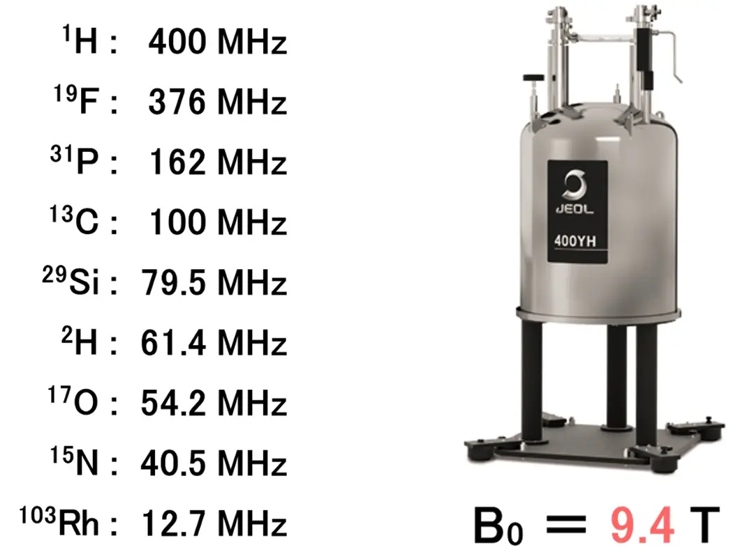
For example, in case of static magnetic field strength of 9.4 tesla (T), 1H nuclei resonates at
400 MHz.
Therefore, an NMR instrument with a magnetic field strength of 9.4 T is generally referred to as a 400 MHz
NMR spectrometer.
However, in multinuclear NMR, nuclei other than 1H resonate at frequencies different from that of
1H, to observe various nuclei such as 13C.
The resonance frequency ν0 is expressed in the relational expression as shown below.
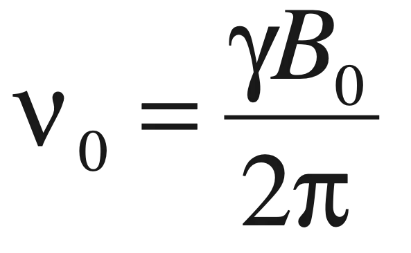
ν0 : Resonance frequency
γ : Gyromagnetic ratio
B0 : Static magnetic field strength
As known from the formula, the resonance frequency ν0 has the relationship that is,
- proportional to static magnetic field strength B0
- proportional to gyromagnetic ratio γ, which is the constant unique to each nucleus.
Gyromagnetic ratio γ is the ratio expressing the speed of precession of each spin in the magnetic field. It is a constant specific to each atomic nucleus. Therefore, this value never changes.
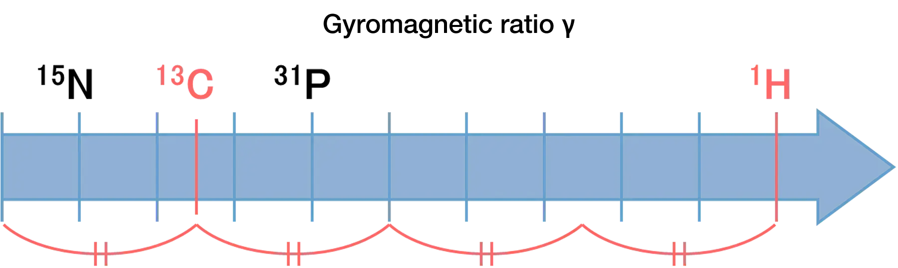
For instance, as illustrated above, the gyromagnetic ratio of 13C is about 1/4 that of
1H. Therefore, the resonance frequency of 13C with a 400 MHz NMR instrument is
approximately 100 MHz (100.5 MHz).
In a similar way, other atomic nucleus has its own gyromagnetic ratio, therefore resonates at a characteristic frequency, even if in the same magnetic field. For example, in case of a 400 MHz instrument (static magnetic field strength = 9.4T), each nucleus resonates at the frequency below.
Key factors determining detection sensitivity
One of the major challenges in multinuclear NMR is "low sensitivity". The detection sensitivity of each nuclear spin (theoretical limit) is expressed in the formula below.
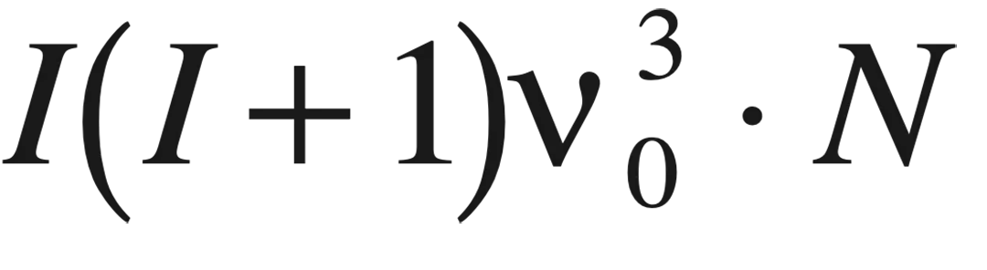
I : Spin quantum number
ν0 : Resonance frequency
N : Nuclear spin concentration (sample volume, natural abundance of isotopes)
The smaller the spin quantum number, the lower the sensitivity. And the lower the resonance frequency, the
lower the sensitivity.
On the other hand, 1H and 19F which have high gyromagnetic ratios and high natural
abundance, and therefore provide relatively high detection sensitivity. Nuclear spin concentration, also a
decisive factor for detection sensitivity, is greatly related with the natural abundance of isotopes other
than the sample volume. For example, in the case of carbon, the natural abundance of 13C is only
1.1%, which results in a corresponding decrease in sensitivity.
The chemical shift range also affects how easily signals can be found. Generally, the greater the atomic
number, the wider the chemical shift range.
For example, the chemical shift range of 1H is about 10-15 ppm. The chemical shift range of
13C is about 200 ppm. For 31P, the observable chemical shift range can extend over several
hundred ppm depending on the chemical environment. The wider the chemical shift range, the more difficult it
is to locate/find the target NMR signals. So, if you widen the observation range blindly, it may be
ineffective. To find the signal, it is recommended to assume the chemical state of the target nuclear spin,
and roughly estimate the position that the signal appears, based on the literature data.
Selecting chemical shift reference standard and points to note
In NMR measurements, selecting the reference standard for calibrating the 0 ppm chemical shift is a critical
step, as it forms the foundation of the entire analysis. For the measurement of 1H and
13C in an organic solvent, TMS (Tetramethylsilane) is commonly used.
For an aqueous sample, DSS (4,4-dimethyl-4-silapentane-1-sulfonic acid) and TSP (Trimethylsilyl propanoic
acid) are selected as standard. This is a common practice.
However, the reference standard used for chemical shift calibration differs in multinuclear NMR, depending on the industry and research field. For example, in 15N NMR, in the organic chemistry field, nitromethane is used as the 0 ppm reference, while in the biochemistry and life science fields, ammonia is often used as the 0 ppm reference.
Here we need to pay attention that there is a chemical shift difference of approx. 400 ppm between
nitromethane and ammonia.
The chemical shift of a target signal can be totally different, depending on the substance used. It is
important to clearly state which substance is defined as 0 ppm in papers and reports.
How to determine pulse width (Nutation experiment)
In multinuclear NMR measurements, it is necessary to know the appropriate 90° pulse width according to the target nucleus to obtain the optimal signal intensity. There are two main methods for obtaining the 90° pulse width.
1. Nutation experiment
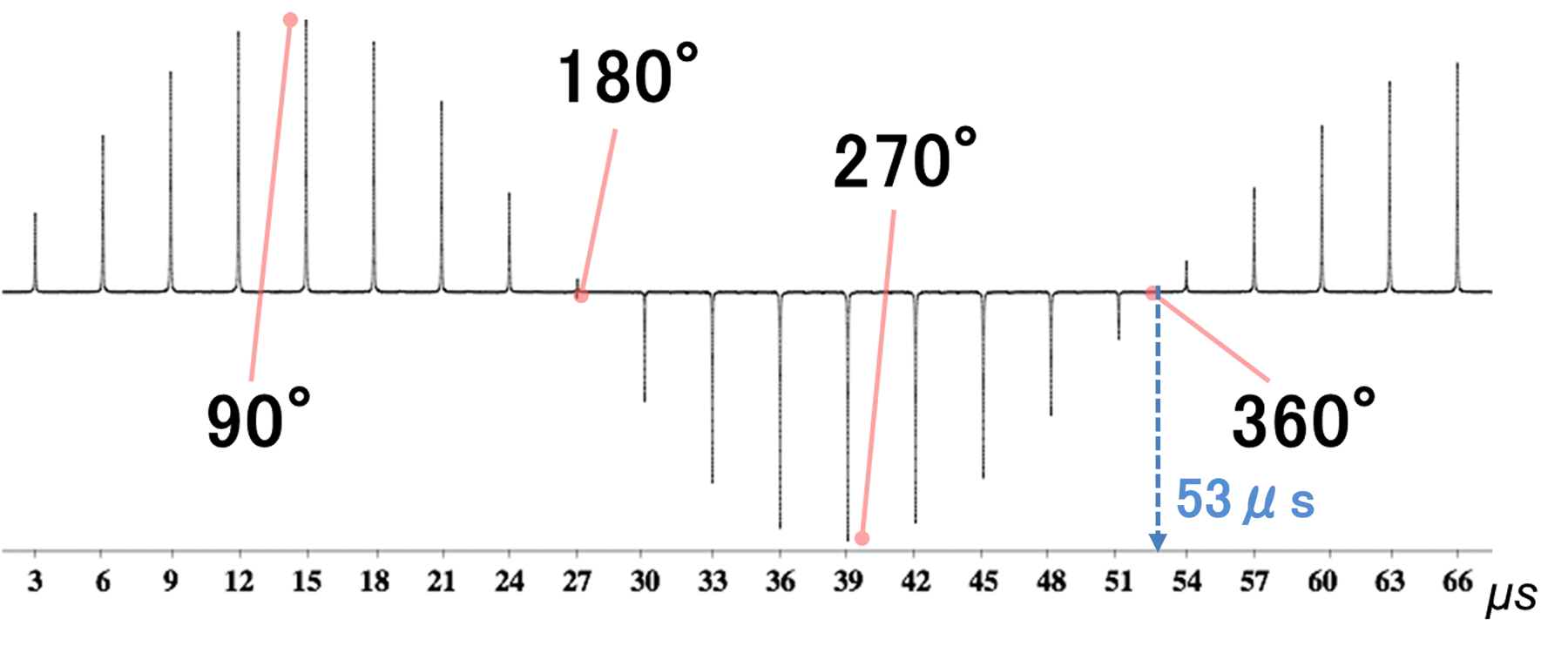
One common method is the nutation experiment. This method involves continuously acquiring spectra while
gradually changing the pulse duration and monitoring the resulting changes in signal intensity.
The signal intensity varies as a sine curve with respect to the pulse width (duration).
- 90° : The signal intensity reaches its maximum in the positive direction.
- 180° : The signal is canceled out, resulting in zero intensity (null point).
- 270° : The signal intensity reaches its maximum in the negative direction.
- 360° : The signal again becomes zero (null point).
In Nutation experiment, the maxima at 90° and 270° are often relatively broad, making it difficult to determine the exact time. Identifying the 360° pulse is generally considered a more reliable and effective approach. In the example shown above, 53 µs corresponds to the 360° pulse width. Therefore, one quarter of this value, 13.25 µs, corresponds to the appropriate 90° pulse width.
Why use the 360° null point instead of the 180° null point?
180° and 360° are both points where the signal disappears (null point), but the 180° null
point tends to be influenced by the "relaxation time" and may be less reliable.
Therefore, in general, the most correct way is to search for the 360° pulse and calculate the 90°
pulse width from that.
2.Estimating the pulse width by calculation
If the signal is too weak to perform the nutation experiment, or if it is impossible to assume the pulse width, it is possible to estimate the pulse width by calculation using data for each known nucleus.
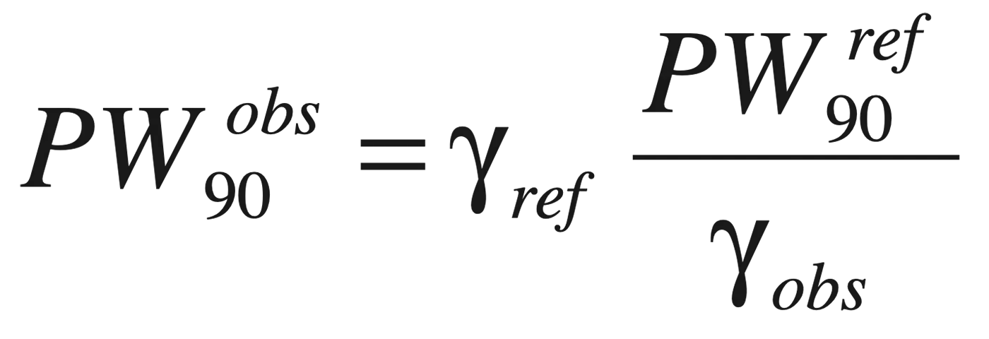
γ (gyromagnetic ratio) can be replaced with ν (resonance frequency)
Principle : this is to utilize the nature of the pulse width that is almost in inverse proportion to the resonance frequency (gyromagnetic ratio γ) of its nucleus.
How to calculate: By using 90° pulse width of known nucleus (i.e. carbon) as the reference, the pulse
width is calculated from the frequency ratio against the target nucleus.
It is more accurate to refer to the 90° pulse width of the nucleus which has a close resonance frequency
to the target nucleus and calculate the 90° pulse width of the target nucleus.
Pulse width and excitation bandwidth
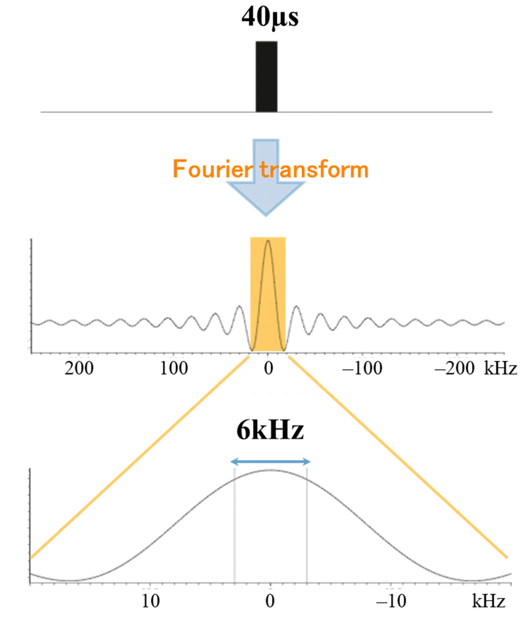
Excitation bandwidth is the range of frequencies that can be adequately excited by an RF pulse. In NMR, it is important if the excitation bandwidth uniformly covers the chemical shift range that you want to observe.
The relationship between the pulse width and excitation bandwidth is as below.

For example, if the 90° pulse width value is 40 µs, the corresponding RF field strength will
be approximately 6 kHz (6250 Hz).
In case of 600 MHz instrument, the excitation bandwidth of 6 kHz is equivalent to a chemical shift
range: 10 ppm.
(Refer to the column No. 1 for calculation of ppm)
However, as can be seen from the waveform of the actual applied pulse shown in the figure, it becomes clear that a 90° pulse width of 40 µs or less is required to sufficiently excite a spectral range of 10 ppm.
Contact Us
JEOL offers a wide range of NMR instruments, probes, and applications for multinuclear NMR measurements.
If you are interested in measurements of low-sensitivity nuclei, analysis of quadrupole nuclei, and a high
sensitivity measurement method, please feel free to contact us. Our experienced application specialists
provide comprehensive support from optimizing measurement conditions to data analysis.

JEOL Ltd.
Since its foundation in 1949, JEOL has been committed to the development
of cutting-edge scientific and metrology instruments, industrial and medical equipment.
Today,
many of our products are used throughout the world and we are highly regarded as a truly global
company.
Aiming to be a 'top niche company that supports science and technology around the
world', we will continue to respond precisely to the increasingly sophisticated and diverse needs
of our customers.
Contacts
JEOL provides a variety of support services to ensure that our customers can use our products with peace of mind.
Please feel free to contact us.