

Exploring "the World of Soft Materials" with Cryo-EM
INTERVIEW 21
Cell Structural Physiology, Graduate School of Science
University of
Hyogo
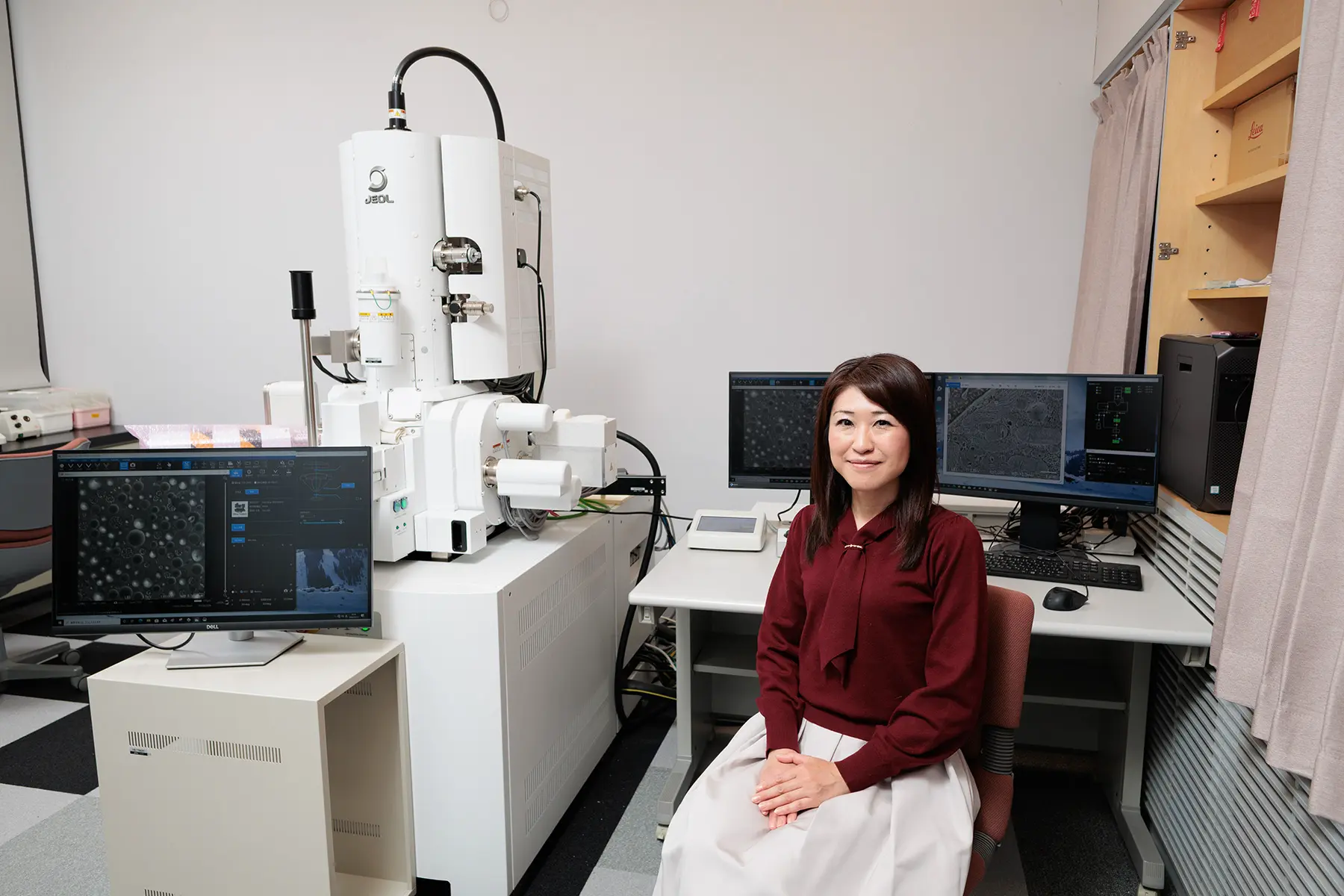
Dr. Yuri Nishino
"We want to observe the state of emulsions". In 2012, a food manufacturer approached Professor Atsuo Miyazawa
at the University of Hyogo with this request and an observation using cryo electron microscopy (Cryo-EM) had
been conducted at the University of Hyogo. Associate Professor Yuri Nishino (then a researcher at RIKEN), a
member of the research group that conducted the observations, recalls that this experience marked the
beginning of her subsequent involvement in soft material observation.
Later, when Dr. Nishino began attending academic conferences related to soft materials, she frequently heard
comments such as, "We want to observe the microstructures in emulsions and slurries, but there is no method
to do so". These experiences made her realize that the Cryo-EM technique observing specimens in a frozen state was not widely known, regardless of whether the instrument is available or not.
For Dr. Nishino, who studies biology, Cryo-EM was already a familiar instrument. Her laboratory had
introduced the cryo-transmission electron microscope (Cryo-TEM) early on, followed by the cryo-scanning
electron microscope (Cryo-SEM).
Cryo-EM became widely recognized after the 2017 Nobel Prize in Chemistry brought the technique into the
spotlight.
By the way, an emulsion is a type of emulsified liquid. Substances in which oil droplets are dispersed in water (or an aqueous
solution) such as milk, mayonnaise, cosmetic lotions, liquid foundations, and hair treatments are called oil-in-water (O/W) emulsions.
Conversely, substances in which aqueous components are dispersed within an oil phase -such as butter, margarine, and moisturizing creams- are called water-in-oil (W/O) emulsions. Both are materials commonly handled in the food and cosmetics industries.
A slurry, on the other hand, is a dispersion of fine solid particles in a liquid, typically with particle sizes between 0.1 µm to 1
mm. It is a materials extensively utilized in battery electrodes, inks, paints, and more.
Are the particles aggregated?
Is the particle size uniform?
Do the structures change over time?
Even when researchers want to observe these structures, because these materials are in a liquid state, conventional electron microscopy, which requires
samples to be dried, cannot capture them in their native states.
"It's the first time, we have seen this!" Voices of joy were raised.
"Then, why don't we show the Cryo-SEM images". Dr. Nishino and her colleagues took some images of
emulsions using Cryo-EM and presented the results. The voices of joy have arisen, "We've never seen this
before!" "This is incredible."
These responses helped spread interest in Cryo-EM for soft material observation. Among the most
enthusiastic were automotive companies researching fuel cells.
In fuel cells, the catalyst layer where electrochemical reactions such as hydrogen oxidation and oxygen
reduction occur is manufactured by coating and drying an ink. Until then, the microstructure and
dispersion of particles within the ink could not be directly observed. Cryo-EM made it possible to
visualize the state of catalyst particles (platinum nanoparticles supported on carbon substrate) within
the ink, contributing to improvements in manufacturing processes.
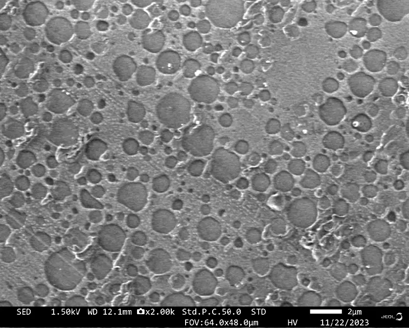
Fresh cream observed by Cryo-SEM (JSM-IT800)
When a cryo-ultramicrotomed surface is observed without coating, oil droplets appear with darker
contrast compared to the aqueous phase.
Moreover, Dr. Nishino's team presented these results at academic conferences, which led to a gradual increase of collaborative research. Some companies even began introducing Cryo EM themselves.
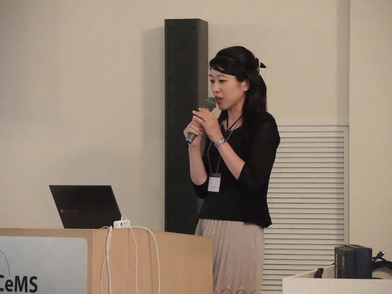
Lecture at the electron microscopy summer school
Accumulating New Know-Hows
As more companies became interested in Cryo-EM and Dr. Nishino began to support them, she faced a new challenge: She needed to explore sample preparation methods suitable for observing each of the various materials. There is not a single method to prepare a specimen.
Not all samples contain water. Some use organic solvents such as alcohol, while others are oil-based, or
contain no water at all. Each requires a different freezing method. Sensitivity to electron beam damage
also varies, and some samples exhibit unexpected physical behaviors depending on the observation method.
In biological samples, liquid components are mostly water, and established know-how exists. But for
non-biological soft materials, almost nothing was known about their behavior under cryogenic vacuum
conditions. Dr. Nishino began exploring these methods through trial and error.
Fortunately, collaborative research with various companies and technical support through the
university's open facility system allowed her to accumulate knowledge on sample preparation, handling,
and observation techniques. She shares this know-how through seminars and workshops, further increasing
industry interest in Cryo-EM.
A New Cryo-SEM with Surprising Advances
As mentioned earlier, the University of Hyogo is equipped with both the Cryo-TEM and Cryo-SEM. The choice of which instrument to use seems to depend
on the size of the target structure:
Cryo-SEM for structures from several hundred nanometers to several micrometers
Cryo-TEM for structures from a few nanometers to several tens of nanometers.
At present, Cryo-SEM is said to be overwhelmingly used for the observation of soft materials such as emulsions and slurries.
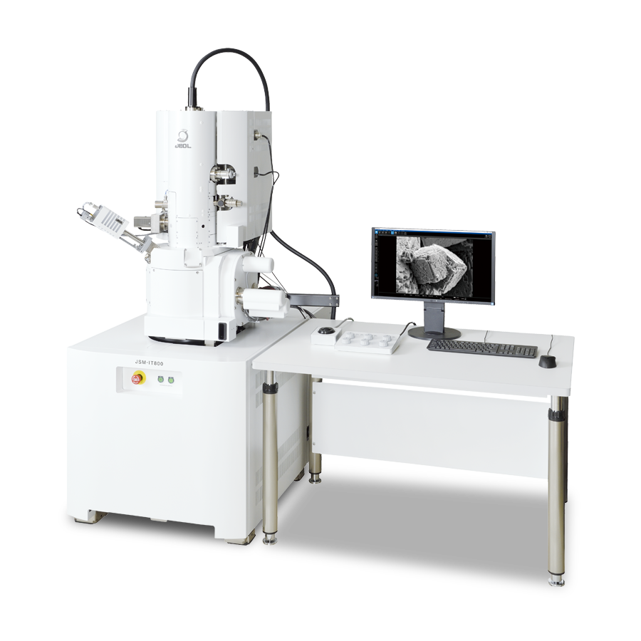
In 2024, the university introduced a new SEM: JEOL JSM-IT800 Schottky Field Emission SEM Equipped with a
cryo-stage and docking port for a vacuum cryo-transfer shuttle, it can be operated as a Cryo-SEM.
Compared with the previous SEM introduced more than a decade ago, the improvements were striking to Dr. Nishino.
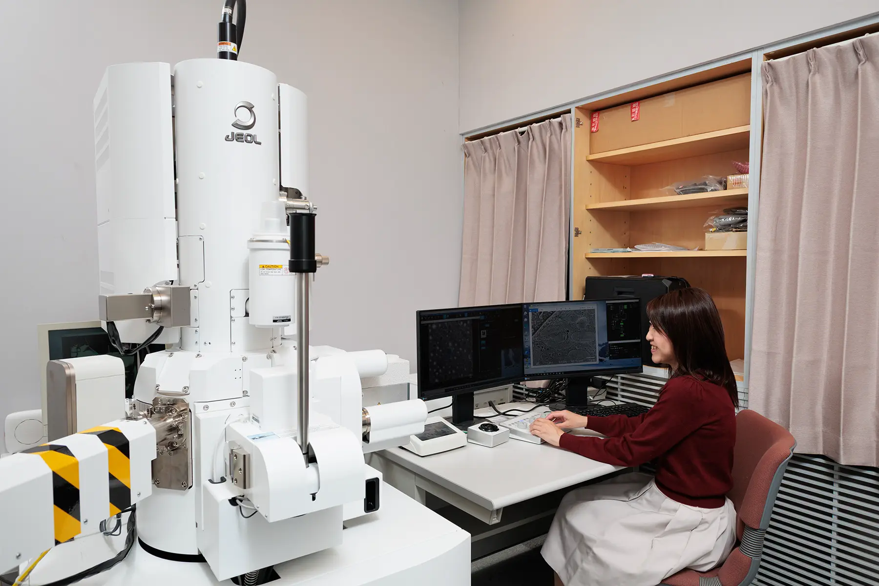
JSM-IT800 Schottky Field Emission Scanning Electron Microscope with cryo
stage
used in observation of soft materials
"The first thing that amazed me was the sharpness of the images," said Dr. Nishino. "The sharpness becomes especially apparent at high magnification.".
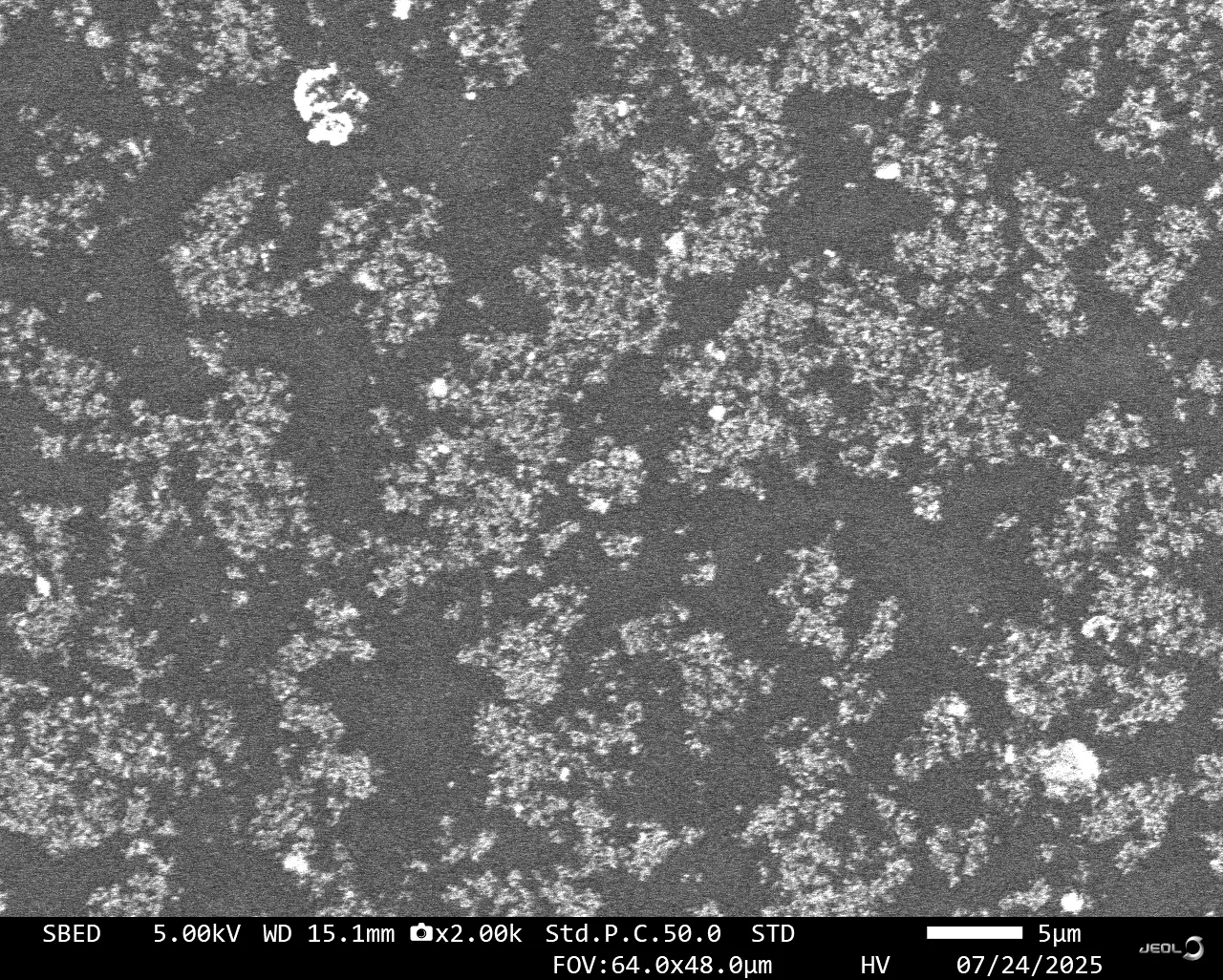
Backscattered electron image of fuel cell ink by Cryo-SEM(JSM-IT800)
While conventional method visualized the structure of the catalyst layer after drying, Cryo-SEM, enables the direct visualization of the particle dispersion in the ink before coating & drying.
She was also impressed by the drift correction function.
With Cryo-SEM, image fluctuations (drifts) often occur. As conductive metal coating is not applied,
specimens are charged up with continuous irradiation of electron beams, causing drifts of detection
images. With previous models, the drifts were troublesome. However, recent software-based corrections now
produce stable images without drifts in most cases, according to Dr. Nishino.
Improved sensitivity of a backscattered electron (BSE) detector is another welcome progress. "When we
tried to observe a BSE image in live mode, it was almost not visible. But now they appear clearly." (Dr.
Nishino). In addition, the image of the BSE detector is adequately clear, it is now sufficient to use a
BSE image as an observation result instead of a secondary electron image. Because BSE images reflect a
compositional contrast, they can be used for image analysis such as binarization and area ratio
calculations.
The Future of soft material research and Cryo-EM
Although the use of cryo-EM in soft materials research has expanded significantly, Dr. Nishino is convinced that "there is still considerable potential for further growth".
Food, cosmetics, and battery industries are already adopting the technique, but many other fields dealing
with soft materials remain unaware of Cryo-EM's potential.
Awareness of cryo-EM remains limited across many industries and fields.
Dr. Nishino and her team plan to continue outreach to these industries. The sharp images produced by the
newly introduced Cryo-SEM may likely serve as powerful tools in their presentations.
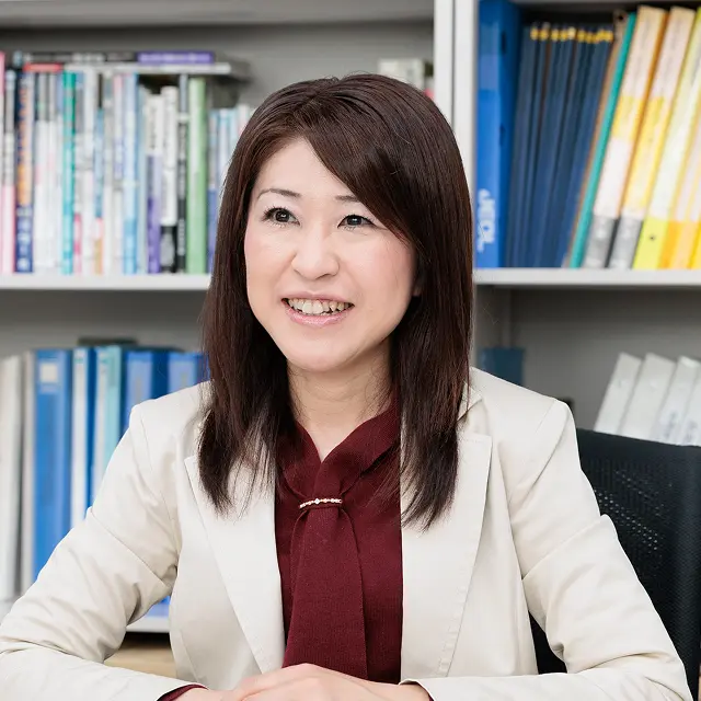
Dr. Yuri Nishino
Associate Professor, Cell Structural Physiology, Department of Life
Science
Graduate School of Science, University of Hyogo
Researcher, RIKEN SPring-8 Center since 2001
Project Assistant Professor Graduate School of Science, University of Hyogo since 2013
Associate Professor since 2025